Osteoporotic Vertebral Fractures
Compression and burst fractures: recognition, imaging, treatment, and the fracture that predicts the next one
- Audience
- Orthopaedic residents, Emergency physicians, Hospitalists, PAs / APPs
- Duration
- 50 minutes (paced estimate ~61 min)
- Slides
- 60 across 10 sections
- References
- 152 verified · 10 attributed images
- Presenter
- Aaron Wey, MD, FAAOS, FACS
- Version
- 1.2 · ready
- Created
- 2026-09-26
- Last updated
- 2026-09-27
- Literature reviewed through
- September 2026
Research record: research/osteoporotic-vertebral-fractures/
Learning objectives
- Recognize an osteoporotic vertebral fracture as a sentinel event: its epidemiology, its effect on mortality and function, and the risk of the next fracture.
- Image correctly: radiographs, when to obtain MRI (acuity, posterior wall, edema) or CT, and how to separate a benign osteoporotic collapse from a malignant one.
- Classify the fracture with the Genant grade, TLICS, the AOSpine thoracolumbar system and the DGOU OF type, know what each is for and where the trauma systems undershoot in osteoporotic bone, and identify the burst fracture, the Kümmell lesion and the neurological deficit that change management.
- Deliver evidence-based nonoperative care: analgesia, early mobilisation, the actual evidence for bracing, and follow-up for progressive collapse.
- State what the randomised trials say about vertebroplasty and kyphoplasty, who is a reasonable candidate, and what the guidelines conclude.
- Know what the trauma trials say about the intact burst fracture, recognize indications for surgery in osteoporotic burst fractures and collapse with neurological deficit, and explain why short-segment fixation fails in osteoporotic bone and how the anterior column is supported.
- Start bone-health treatment: DXA, FRAX, laboratory work-up, the antiresorptive and anabolic agents by name with their trial efficacy, sequencing and safety, and referral to a fracture liaison service.
Osteoporotic Vertebral Fractures
Compression and burst fractures: recognition, imaging, treatment, and the fracture that predicts the next one
Outline
- 1.Introductionslide 1
- 2.Why it mattersslide 4
- 3.Mechanism & classificationslide 8
- 4.Recognition & imagingslide 20
- 5.Nonoperative careslide 28
- 6.Vertebral augmentationslide 32
- 7.Burst fractures & surgeryslide 39
- 8.Bone healthslide 46
- 9.Casesslide 53
- 10.Summaryslide 57
All slides
Osteoporotic Vertebral Fractures
Compression and burst fractures: recognition, imaging, treatment, and the fracture that predicts the next one
A 78-year-old woman lifts a bag of groceries and feels something give in her back. The radiology report says “compression deformity of L1, age indeterminate.”
She is discharged with analgesics. Nobody orders a DXA. Eleven months later she is back with T12.
Learning objectives
- Treat the vertebral fracture as a sentinel event: epidemiology, mortality, and the risk of the next fracture
- Image correctly: standing radiographs, MRI for acuity and the posterior wall, CT for the burst, and how to spot malignancy
- Classify the fracture (Genant, OF, AOSpine) and identify the burst, the Kümmell lesion, and the deficit that change management
- Deliver evidence-based nonoperative care and know what bracing actually does
- State what the randomized trials say about vertebroplasty and kyphoplasty, and who is a reasonable candidate
- Recognize surgical indications in osteoporotic burst fractures and the principles of fixation in weak bone
- Start bone-health treatment before the patient leaves your care
Why it matters
Common, underdiagnosed, and a predictor of death and of the next fracture
The most common osteoporotic fracture
One in five patients with a new vertebral fracture has another vertebral fracture within a year.
19.2% (95% CI 13.6–24.8) in 2,725 placebo-arm women; a prevalent fracture raised the risk 5-fold. Lindsay 2001 Klotzbuecher 2000
Not a benign injury
Mechanism and classification
What breaks, how much, and which fractures are not the simple kind
Why the vertebra fails
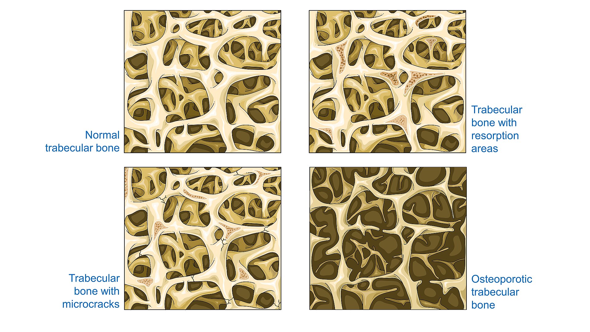
- Osteoporosis thins and disconnects the trabecular network; the vertebral body is mostly trabecular bone and fails under axial load and flexion
- Thoracolumbar junction is the commonest site: L1 in 19% of one inpatient series, with T12 and L2 close behind Schnake 2022
- Minimal or no trauma: 83% of clinical fractures follow moderate or no trauma; half of hospitalised patients recall no injury Cooper 1992 Schnake 2022
- Anterior wedge is typical; end-plate failure gives biconcave and crush patterns
- Some fractures keep moving: 44% of vertebroplasty candidates showed dynamic mobility between standing and supine films McKiernan 2003
- Progression of deformity is linked to a T-score below −3 Schnake 2022
Genant semiquantitative grading
Wedge, biconcave and crush morphologies by degree of height loss. Original diagram after Genant 1993. Genant 1993
- Grade 1, mild: 20–25% loss of anterior, middle or posterior height
- Grade 2, moderate: 25–40% loss
- Grade 3, severe: more than 40% loss; multiple grade 2–3 fractures define severe osteoporosis in every guideline
The OF classification
AO Spine–DGOU osteoporotic fracture types. Original diagram after Schnake 2018. Schnake 2018 Scherer 2025
Four generations of thoracolumbar injury classification
Thoracolumbar Injury Classification and Severity Score
Score the three domains and add them. Original diagram after Vaccaro 2005. Vaccaro 2005 Petfield 2011
- Morphology on radiographs, CT and MRI: compression 1, burst 2, translation or rotation 3, distraction 4; only the most severe pattern scores
- PLC: supraspinous and interspinous ligaments, ligamentum flavum, facet capsules. Palpate for a gap; look for spinous process splaying, facet subluxation and STIR edema
- Neurology from the ISNCSCI examination; incomplete cord, conus and cauda equina injuries score highest because they have the most to gain
- ≤ 3 nonoperative, ≥ 5 operative, 4 is the surgeon's call; the score matched actual treatment in 96% of cases Joaquim 2011
The AOSpine thoracolumbar classification
Morphology types, neurological grade and modifiers. Original diagram after Vaccaro 2013. Vaccaro 2013
Confounders TLICS and AOSpine cannot score
The confounders
- Elderly: mortality after geriatric trauma rises about 7% per year over 65; comorbidity may argue against surgery the score recommends
- Osteoporosis: screws pull out; cement augmentation, hooks or bands, or no surgery if fixation cannot hold
- Cannot mobilise or tolerate a brace: the threshold for surgery falls to avoid bed-rest complications
- Ankylosed spine (AS, DISH): long lever arms, transverse fractures hidden on radiographs; low threshold for CT, MRI and fixation
- Obesity: braces are ineffective; the TLICS 2 burst may need short-segment fixation instead
- Polytrauma: operate to mobilise a patient who cannot lie in a brace
What the OF classification adds
- Describes the low-energy failure modes trauma systems ignore: edema-only OF1, collapse, pincer and Kümmell OF4
- Expects progression: the type is upgraded on standing follow-up films
- An OF-type-based score (< 6 nonoperative, > 6 surgical) that weighs clinical factors alongside morphology
- Principles for weak bone: stability over motion, cement-augmented or high-purchase screws, restore the patient's own sagittal profile
- Minimally invasive and augmentation-based options as first-line surgery
Textbook chapters reviewed: Vaccaro et al., Spine and Spinal Cord Trauma (Thieme, 2011). Williams 2011 Schmidt 2011 Blattert 2018
A 76-year-old woman falls from standing. CT shows an L1 fracture involving the superior end plate and the posterior wall with 25% canal retropulsion and no facet widening. MRI: intact posterior ligamentous complex. She is neurologically intact. How would you classify and score this injury?
- ATLICS 5 (burst 2 + PLC indeterminate 2 + intact 0 + osteoporosis 1): operative
- BTLICS 2 (burst 2, PLC intact 0, neurology intact 0); AOSpine A3 N0 M2; OF3: nonoperative by the trauma scores, with close standing-film follow-up because of the bone
- CTLICS 4 (burst 2 + PLC intact 0 + retropulsion 2): surgeon's judgement
- DAOSpine A1, TLICS 1: discharge with analgesia and no follow-up imaging
Which classification for which question
| System | Answers | Use it when |
|---|---|---|
| Genant Genant 1993 | How much height is lost, and how many vertebrae | Reading radiographs, defining severe osteoporosis, choosing anabolic therapy |
| Denis three-column Denis 1983 | Which columns have failed | Bedside vocabulary: compression vs burst vs flexion-distraction |
| TLICS Vaccaro 2005 | Morphology + PLC + neurology as a score, and the approach | Communicating any thoracolumbar injury to the surgeon in one line |
| AOSpine thoracolumbar Vaccaro 2013 Kepler 2016 | A0–A4 / B / C morphology, N grade, M2 flags osteoporosis | Registries, research and high-energy or ankylosed-spine injuries |
| OF classification Schnake 2018 Blattert 2018 | Posterior wall, collapse, instability, and progression over time | Deciding between nonoperative care, augmentation and instrumentation in osteoporotic bone |
In every system the posterior wall and the neurological examination are the pivots; only the OF type is designed to be upgraded on follow-up imaging.
Nonunion, clefts and Kümmell disease
- 13.5% of conservatively treated fractures were nonunited at 6 months in a prospective series of 350 patients Tsujio 2011
- Predictors of nonunion: thoracolumbar level, middle-column injury, and a confined high or diffuse low T2 signal on the initial MRI Tsujio 2011
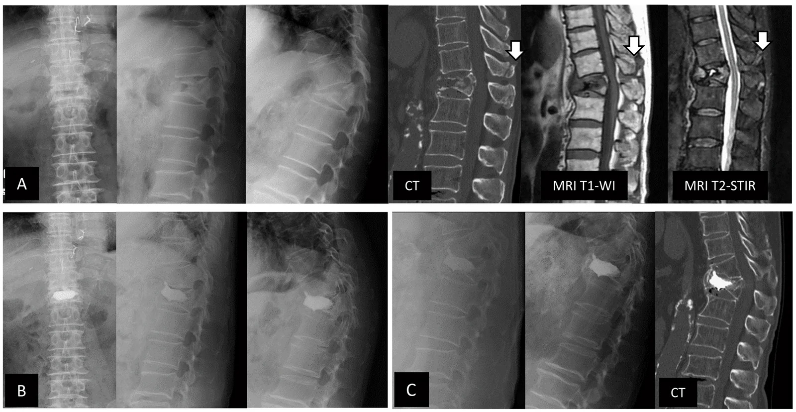
- Intravertebral cleft (vacuum) = mobile, unhealed fracture; present in every mobile fracture and in 79% of delayed collapses before a neurological deficit appeared McKiernan 2003 Ito 2002
- Kümmell disease: delayed post-traumatic avascular collapse of the vertebral body, weeks to months after a seemingly minor fracture Freedman 2009
- Predictors of collapse and deficit: cleft, T1 total or T2 wide hypointense patterns, retropulsion over 42% of the canal, or more than 15° of dynamic wedge change Muratore 2018
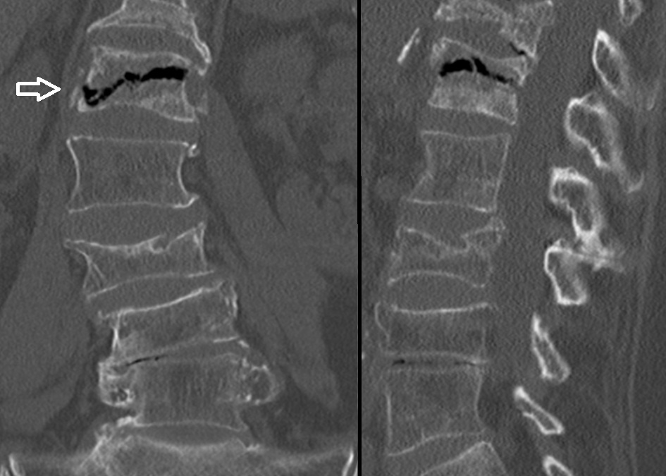
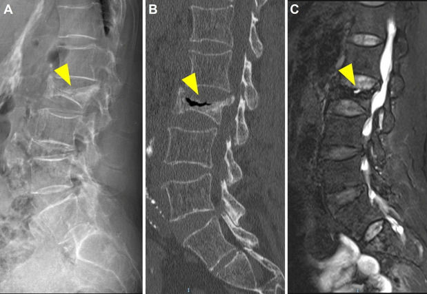
Kümmell disease
Recognition and imaging
Is it acute, is the posterior wall intact, and is it osteoporosis at all?
What the patient tells you
- Acute, localised midline thoracolumbar pain after minimal or no trauma; worse standing and turning in bed, eased lying flat
- Often no neurological symptoms; radicular pain occasionally; ask specifically about leg weakness, numbness and bladder function
- Loss of height, progressive kyphosis, early satiety and shortness of breath in chronic or multiple fractures
- Percussion tenderness over the spinous process at the fractured level; a painful gap or step suggests a more severe injury
- Risk profile: age, prior fragility fracture, glucocorticoids, low weight, smoking, alcohol, and the medications and diseases that weaken bone
- Red flags for another diagnosis: fever, weight loss, night pain, known malignancy, age under 50, pain above T5, or a deficit
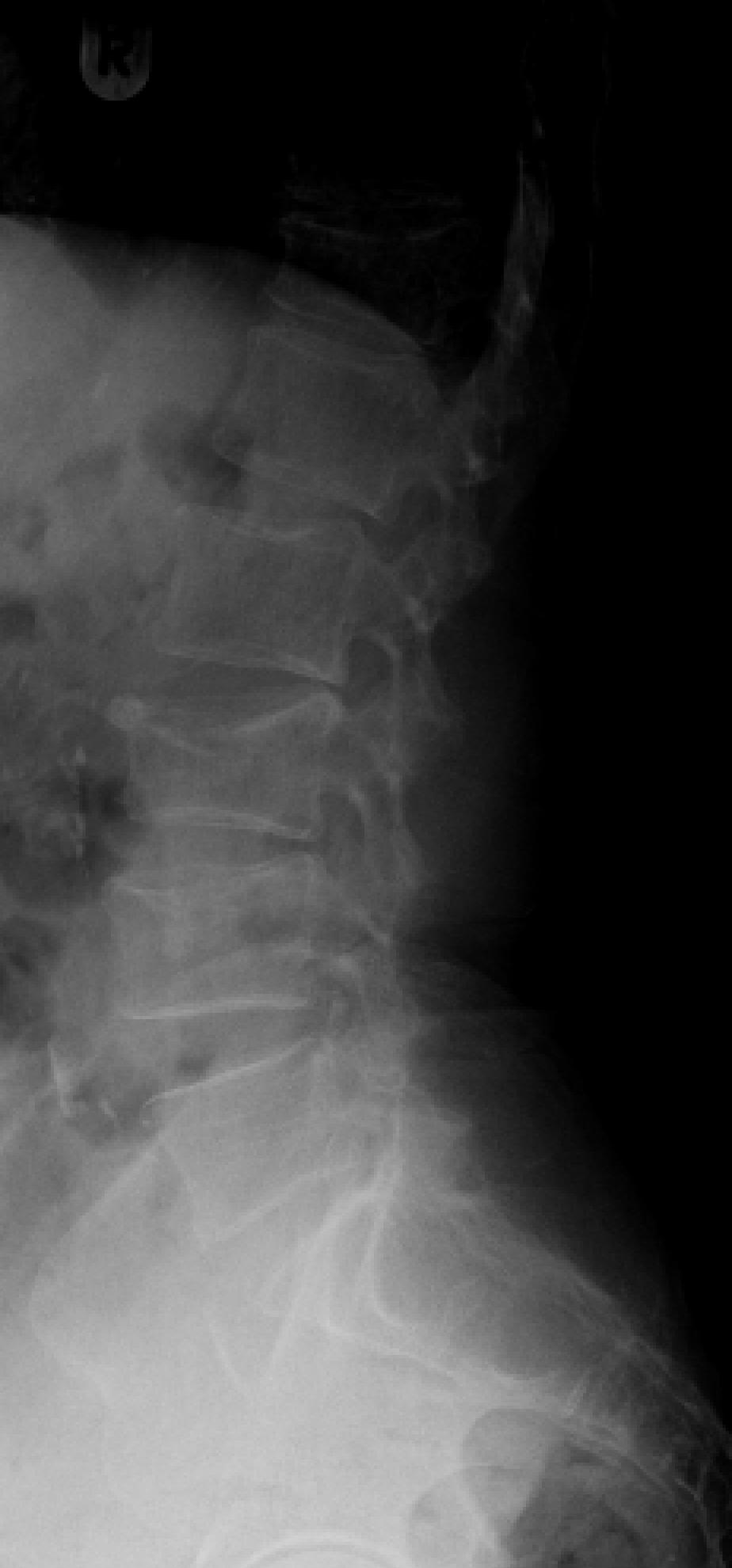
Not every collapsed vertebra is osteoporosis
Osteoporotic
- Older patient, minimal trauma, thoracolumbar junction
- Band-like edema under the end plate; normal marrow signal elsewhere
- Retropulsed fragment, intact posterior cortex line
- Often multiple healed fractures
- Normal calcium, alkaline phosphatase, blood count
Malignant
- Known cancer, weight loss, night pain, atypical level
- Convex posterior cortex, pedicle or posterior element signal change
- Epidural or paraspinal soft-tissue mass, other lesions
- Diffuse marrow replacement on T1
- Myeloma: anaemia, hypercalcaemia, protein electrophoresis
Other
- Infection: disc-centred, end-plate destruction, fever, raised CRP
- High-energy trauma: unstable patterns regardless of bone quality
- Kümmell / nonunion: cleft, delayed collapse
- Ankylosed spine: transverse fractures through fused segments, grossly unstable
Imaging the suspected osteoporotic fracture
MRI is the preferred advanced study in the expert consensus pathway; bone scan or CT are alternatives when MRI is not possible. Hirsch 2018 Expert 2023
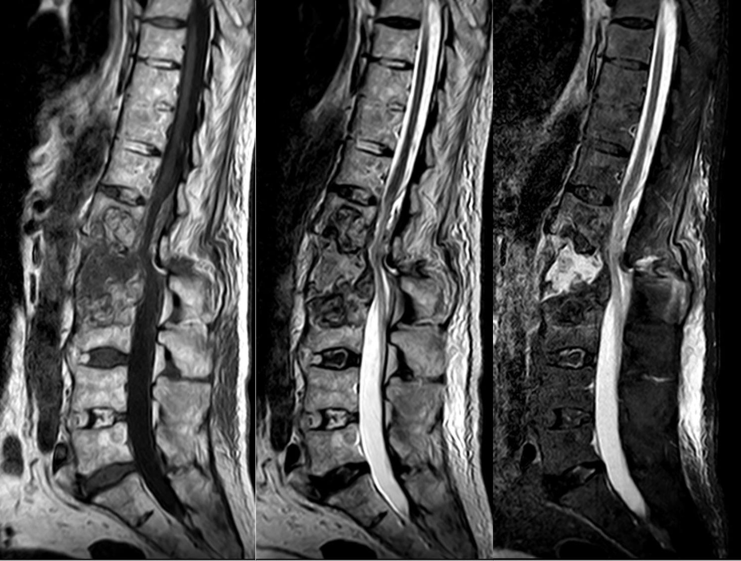
STIR dates the fracture
Benign versus malignant collapse on MRI
| Feature | Favours osteoporotic | Favours malignant |
|---|---|---|
| Posterior cortex | Straight or retropulsed fragment | Convex, bulging posterior border |
| Pedicles and posterior elements | Normal signal | Abnormal signal, involvement |
| Soft tissue | None or thin haematoma | Epidural or paraspinal mass |
| Marrow pattern | Band-like low T1 signal, spared normal marrow | Diffuse replacement, inhomogeneous enhancement |
| Other vertebrae | Multiple compression fractures | Other focal lesions |
| Fluid or cleft | Fluid sign or vacuum cleft | Rare |
Non-fat-suppressed T2 signal alone does not discriminate. When in doubt, biopsy at the time of augmentation or before treatment. Jung 2003 Cuénod 1996
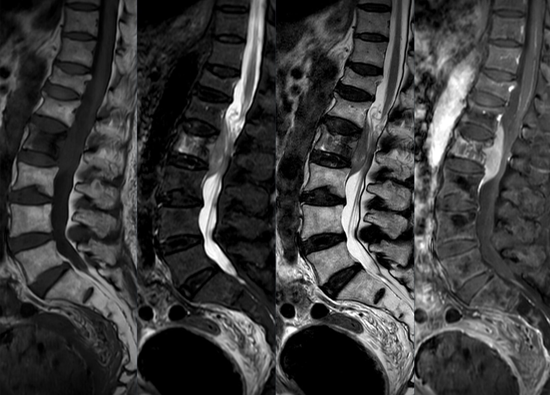
The collapse that is not osteoporosis
An 81-year-old woman presents with 5 days of severe midline pain after sitting down hard. Radiographs show a 30% anterior wedge of T12 that was not present on films from 2 years ago. She is neurologically normal and has no red flags. What imaging, if any, is most appropriate next?
- ANone; the fracture is dated by comparison with the prior films, so treat and repeat standing radiographs in 2–4 weeks
- BMRI now, because every new osteoporotic fracture requires MRI before treatment
- CBone scan to confirm the fracture is acute
- DFlexion–extension radiographs to assess stability
Nonoperative care
Analgesia, mobilisation, and the truth about braces
What nonoperative care actually consists of
- Multimodal analgesia: scheduled paracetamol, short NSAID course if renal function allows, cautious short-acting opioids; calcitonin reduced acute pain in trials (mean −3.4 points at 1 week) Knopp-Sihota 2012
- Early mobilisation with physiotherapy; bed rest beyond a day or two costs bone and muscle and raises thromboembolic and pulmonary risk; mechanical and pharmacological prophylaxis while the patient is immobile (consensus, not trial evidence)
- Safe-movement teaching: hip-hinge, avoid loaded flexion; exercise improves mobility, pain and fear of falling Ponzano 2023; back-extensor strengthening cut vertebral fractures 4.3% → 1.6% over 10 years in one small RCT Sinaki 2002
- Nutrition: early satiety and weight loss are common after thoracic fractures; check intake and vitamin D Ponzano 2023
- Standing radiographs at 2–4, 6 and 12 weeks; escalate if pain fails to settle or the OF type upgrades Blattert 2018
- Expect improvement over weeks: in trials, conservative arms fell from VAS 8 to about 2–4 by 3 months Rousing 2009 Klazen 2010
Does a brace help?
Rigid brace, soft brace, or no brace
| Study | Design | N | Comparison | Key finding | Limitation |
|---|---|---|---|---|---|
Kim 2014 Kim 2014 | RCT, non-inferiority | 60 | No brace vs soft vs rigid, one-level fracture < 3 days | ODI at 12 weeks 36 vs 38 vs 34: no brace non-inferior; pain and compression progression identical | Small; Korean single centre |
Kato 2019 Kato 2019 | Multicentre RCT | 284 | Rigid vs soft brace for 12 weeks | No difference in vertebral compression at 48 weeks, pain, or quality of life | No no-brace arm |
Pfeifer 2004 Pfeifer 2004 | RCT | 62 | Spinomed orthosis 6 months vs none, chronic kyphosis | Back-extensor strength +73%, kyphosis −11%, pain −38% | Chronic patients; unblinded; industry device |
Rzewuska 2015 Rzewuska 2015 | Systematic review | 5 RCTs, 350 | Analgesics and orthoses vs control | Orthoses improved medium-term pain and disability vs nothing (SMD −1.5) | Very low to low quality evidence |
Pieroh (DGOU) 2023 Pieroh 2023 | Systematic review | 13 studies | Orthosis types and no orthosis | No superiority of any orthosis; where compared, non-rigid favoured | Heterogeneous, mostly low quality |
Two randomised trials show a rigid brace is no better than a soft brace, and no brace is non-inferior to either for pain, function and deformity. Orthoses may help some patients feel safer and, in chronic kyphosis, strengthen extensors.
Do not prescribe a rigid TLSO by reflex. Offer a soft or semi-rigid orthosis for comfort and confidence if the patient wants one, mobilise early, and reserve rigid bracing for the surgeon's specific plan.
Predictors and signs of conservative failure
On the initial imaging
- Thoracolumbar junction level
- Middle-column (posterior wall) involvement
- Confined high or diffuse low T2 signal; T1 total or T2 wide hypointense patterns
- Intravertebral cleft or fluid sign
- Retropulsion over 42% of the canal
On follow-up
- Pain not settling by 4–6 weeks despite adequate care
- Progressive height loss or kyphosis on standing films; OF type upgraded
- More than 15° change in the wedge angle between positions
- New neurological symptoms or signs
- Immobility, inability to mobilise despite analgesia
Predictors from prospective and pooled data. Tsujio 2011 Muratore 2018 Ito 2002 Blattert 2018
Vertebral augmentation
Vertebroplasty, kyphoplasty, and a decade of contradictory trials
Vertebroplasty and balloon kyphoplasty
Transpedicular approach under fluoroscopy. Original diagram.
- Both use a transpedicular or extrapedicular cannula placed under fluoroscopy, usually under sedation or general anaesthesia
- Vertebroplasty injects low-viscosity cement directly into the fractured trabecular bone under pressure
- Kyphoplasty first inflates a balloon to create a cavity and partially restore height, then fills it with viscous cement at low pressure
- Implantable devices (titanium jack, stents) aim for more durable height restoration; a randomized trial found the titanium device non-inferior to kyphoplasty with fewer adjacent fractures Noriega 2019
What the randomized trials found
Vertebroplasty or kyphoplasty versus sham or conservative care
| Study | Design | N | Comparison | Key finding | Limitation |
|---|---|---|---|---|---|
Buchbinder 2009 Buchbinder 2009 | Sham-controlled RCT | 78 | Vertebroplasty vs sham, < 12 months | No difference at any time point; 3-month pain −2.6 vs −1.9 | Small; long symptom duration allowed |
INVEST (Kallmes) 2009 Kallmes 2009 | Sham-controlled RCT | 131 | Vertebroplasty vs simulated procedure | No difference in RDQ or pain at 1 month; 51% vs 13% crossed over | Sham included periosteal anaesthetic |
Vertos II (Klazen) 2010 Klazen 2010 | Open-label RCT | 202 | Vertebroplasty vs conservative, ≤ 6 weeks, MRI edema, VAS ≥ 5 | VAS difference 2.6 at 1 month, 2.0 at 1 year favouring vertebroplasty | Unblinded; 53% of screened had spontaneous relief |
VAPOUR (Clark) 2016 Clark 2016 | Sham-controlled RCT | 120 | Vertebroplasty vs placebo, < 6 weeks, NRS ≥ 7 | NRS < 4 at 14 days: 44% vs 21% | Sham without periosteal anaesthetic; industry grant |
VERTOS IV (Firanescu) 2018 Firanescu 2018 | Sham-controlled RCT | 180 | Vertebroplasty vs sham, acute | VAS differences 0.2–0.45 over 12 months, not significant | Sham included periosteal anaesthetic |
FREE (Wardlaw) 2009 Wardlaw 2009 | Open-label RCT | 300 | Kyphoplasty vs non-surgical care, ≤ 3 months | SF-36 PCS +7.2 vs +2.0 at 1 month; benefit fades by 12 months, pain still better at 2 years | Unblinded; industry funded |
VERTOS V (Carli) 2023 Carli 2023 | RCT vs active control | 80 | Vertebroplasty vs anaesthetic infiltration, chronic > 3 months with edema | 12-month VAS 3.9 vs 5.1; QUALEFFO better; disability not different | Small; control not a true sham |
Against a sham that anaesthetises the periosteum, vertebroplasty has shown no clinically important benefit; against conservative care or a skin-only placebo, it and kyphoplasty relieve pain faster, with the largest effects in severe pain of under 6 weeks. Effects fade by a year.
Augmentation is not routine care. Consider it for a patient with an MRI-confirmed acute fracture, severe pain despite adequate analgesia, and functional failure (cannot mobilise, hospitalised), after an honest discussion of the trials. It is not a treatment for the bone.
What the guidelines conclude
| Body | Year | Position |
|---|---|---|
| AAOS Esses 2011 | 2010 | Strong recommendation against vertebroplasty; weak recommendation that kyphoplasty is an option in neurologically intact patients |
| Cochrane Buchbinder 2018 | 2018 | High- to moderate-quality evidence of no important benefit over sham; harms cannot be excluded |
| ASBMR Task Force Ebeling 2019 | 2019 | No clinically significant benefit of vertebroplasty over placebo; insufficient evidence for kyphoplasty; routine use not supported; inform patients fully |
| Multispecialty pathway (RAND/UCLA) Hirsch 2018 | 2018 | Augmentation appropriate with positive advanced imaging and worsening symptoms or 2–4 unfavourable conditions; time since fracture less relevant |
| ACR Appropriateness Criteria Expert 2023 | 2022 | Scenario-based ratings; MRI preferred before treatment; augmentation may be appropriate for refractory painful acute fractures |
| NASS guideline Cho 2025 | 2025 | 29 evidence-graded questions on diagnosis and treatment; full guideline on the NASS website |
Specialty bodies read the same trials and reach different conclusions; the divergence tracks who wrote the guideline.
Do augmented patients live longer?
Who, when, and what can go wrong
A reasonable candidate
- MRI-confirmed acute or unhealed fracture with edema, at the level that matches the pain
- Severe pain (≥ 7/10) despite adequate analgesia, or inability to mobilise, especially if hospitalised
- Edema and concordant pain matter more than the calendar: recent fractures gave the largest trial effects, but chronic fractures with persistent edema respond, and kyphoplasty beat conservative care at over 12 months in Kasperk's cohort Láinez 2021 Hirsch 2018 Kasperk 2005
- No infection, no coagulopathy, no malignancy without a plan for biopsy, no significant retropulsion with deficit
Complications
- Cement leakage per vertebra: 41% vertebroplasty, 9% kyphoplasty, mostly silent; symptomatic leakage, cord compression, pulmonary cement embolism and osteomyelitis are reported Hulme 2006 Taylor 2007 Buchbinder 2018
- New fractures cluster early and adjacent: 26% within 8 months of kyphoplasty, the early ones all adjacent Fribourg 2004; cement in the disc raises adjacent fracture from 12% to 58% Lin 2004; pooled RCTs show no excess versus conservative care Halvachizadeh 2021
- Kyphoplasty carries a higher rate of subsequent augmentation in Medicare data (9.4% vs 7.9%) Chen 2013
- Not a minor procedure: a frail patient prone with a kyphotic chest, and a surgeon and anaesthetist available for the epidural leak that needs decompression Schmidt 2011
An 84-year-old man with an MRI-confirmed acute L2 wedge fracture (OF2) has been admitted for 6 days. Despite scheduled analgesics and physiotherapy he cannot stand, his pain is 8/10, and he has developed a sacral pressure area. Which statement best reflects the evidence?
- AVertebroplasty is contraindicated because two sham-controlled trials showed no benefit
- BAugmentation is a reasonable option for this patient, with an honest discussion that sham-controlled trials show small or no average benefit but that he fits the subgroup most likely to respond
- CA rigid TLSO should be fitted and he should be observed for a further 4 weeks before any procedure
- DKyphoplasty is indicated because it reduces mortality by 37%
Burst fractures, collapse, and surgery
When the osteoporotic fracture stops being a benign problem
The osteoporotic fracture that is not benign
- Burst fracture: posterior wall involved (OF3, AOSpine A3–A4) or vertebral frame lost (OF4) Schnake 2018 Vaccaro 2013. Retropulsion alone in an intact patient is not an indication: it remodels Wood 2003 Cantor 1993
- Neurological deficit: rare (about 3% of inpatients) but an indication for decompression and stabilisation Schnake 2022 Blattert 2018
- Delayed collapse with a cleft (Kümmell) and progressive kyphosis; retropulsion over 42% of the canal or over 15° dynamic change predicts deficit Muratore 2018
- Distraction or rotation injuries (OF5, AOSpine B or C, TLICS ≥ 5), and any fracture in an ankylosed spine: 67% of AS fractures present with a deficit and 3-month mortality is about 18% Westerveld 2009 Blattert 2018
- Multiple adjacent fractures with sagittal decompensation: the patient who can no longer stand upright
- Painful nonunion that has failed conservative care and is not suitable for augmentation alone
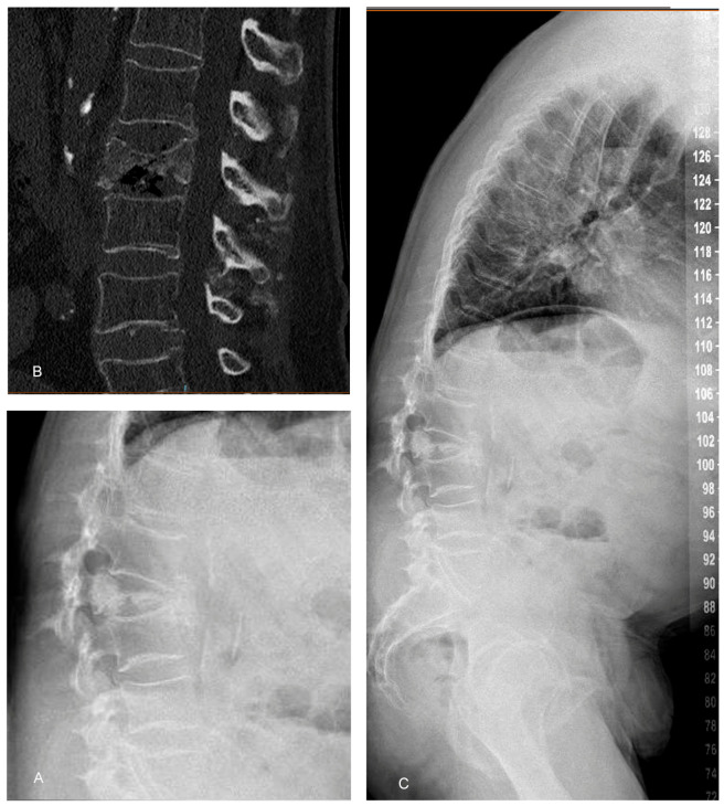
The intact burst fracture: what the trauma trials say
| Study | Design | Result | Message |
|---|---|---|---|
| Wood 2003 Wood 2003 | RCT, 47 stable bursts, no deficit, 44 mo | Kyphosis and canal remodelling equal (39 → 22% vs 34 → 19%); pain and work similar; less disability, fewer complications without surgery | The stable burst is a nonoperative injury |
| Shen 2001 Shen 2001 | 80 patients, brace vs short-segment fixation, 2 y | Surgery: less pain to 3 months, equal by 6; correction lost; braced retropulsion 34 → 15%; charges four-fold | Early comfort, no lasting gain |
| Siebenga 2006 Siebenga 2006 | RCT, 34 patients, 4.3 y | Surgery: less kyphosis, better pain and function, more return to work | The dissenting trial: small, mixed types |
| Bailey 2014 Bailey 2014 | Equivalence RCT, 96 A3 bursts, TLSO vs none | Roland-Morris 6.8 vs 7.7 at 3 months: equivalent; 6 needed surgery | No brace equals a brace, for bursts too |
| Cantor 1993 Cantor 1993 | 18 bursts, intact posterior column, early ambulation | Kyphosis 19 → 20°; retropulsed bone resorbed; no neurological deterioration | The fragment remodels |
Spine Trauma Study Group: bracing 6–12 weeks is reasonable; either approach when surgery is chosen, posterior favoured in the elderly with poor bone. Sekhon 2011
Why short-segment fixation fails in this bone
- Load sharing: comminution, fragment apposition and kyphosis correction predict failure of short-segment posterior fixation; a score of 7 or more needs anterior support McCormack 1994
- Short versus long segment for bursts: 55% implant failure with short-segment fixation, none with long, with the same clinical outcome Tezeren 2005
- Ligamentotaxis reduces the periphery but leaves a central end-plate depression; the disc creeps into it and the kyphosis recurs Oner 1998
- Fill the void and fix the level: cement under the end plate cut screw bending moments 59% Mermelstein 1998; balloon reduction with short-segment screws restored 91% anterior height Verlaan 2005; screws at the fractured level add torsional stiffness Mahar 2007
- Percutaneous hybrid in older patients: kyphoplasty plus short percutaneous screws, kyphosis 16° → 2°, 45 minutes, 70 mL, no implant failure Korovessis 2008; percutaneous halves blood loss with the same 5-year result Wild 2007; open trauma instrumentation infects 10% Rechtine 2001
Surgical strategies in osteoporotic bone
Kyphoplasty alone
- Pooled series of 289 patients with osteoporotic burst fractures: VAS 6.8 → 1.1, kyphosis 22° → 3° Chen 2021
- Requires intact enough posterior wall and no deficit
- Leakage 8–45%, adjacent fracture or relapse common
Posterior fixation ± augmentation
- Short-segment fixation with vertebroplasty of the index level: correction loss 4.6° vs 8.6° without, equal to anterior surgery Uchida 2010
- Cement-augmented or high-purchase screws; stability over motion preservation Blattert 2018
- Long-segment fixation holds kyphosis better in Kümmell disease; short-segment is faster and bloodier-sparing Yu 2022
Anterior or combined
- Anterior strut or Kaneda reconstruction restores the load-bearing column; all recovered neurologically in one series Kanayama 2010
- 34% respiratory complications with anterior surgery; posterior reinforcement needed at BMD < 0.60 g/cm² Sudo 2013
- Posterior closing-wedge osteotomy matches anterior–posterior surgery with less morbidity for fixed kyphosis with deficit Suk 2003
All comparative data are retrospective; no randomised trial exists for surgery in osteoporotic burst fractures with deficit.
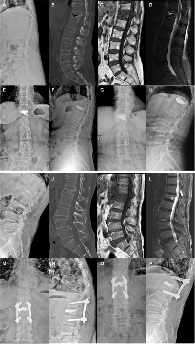
Two answers to the same problem
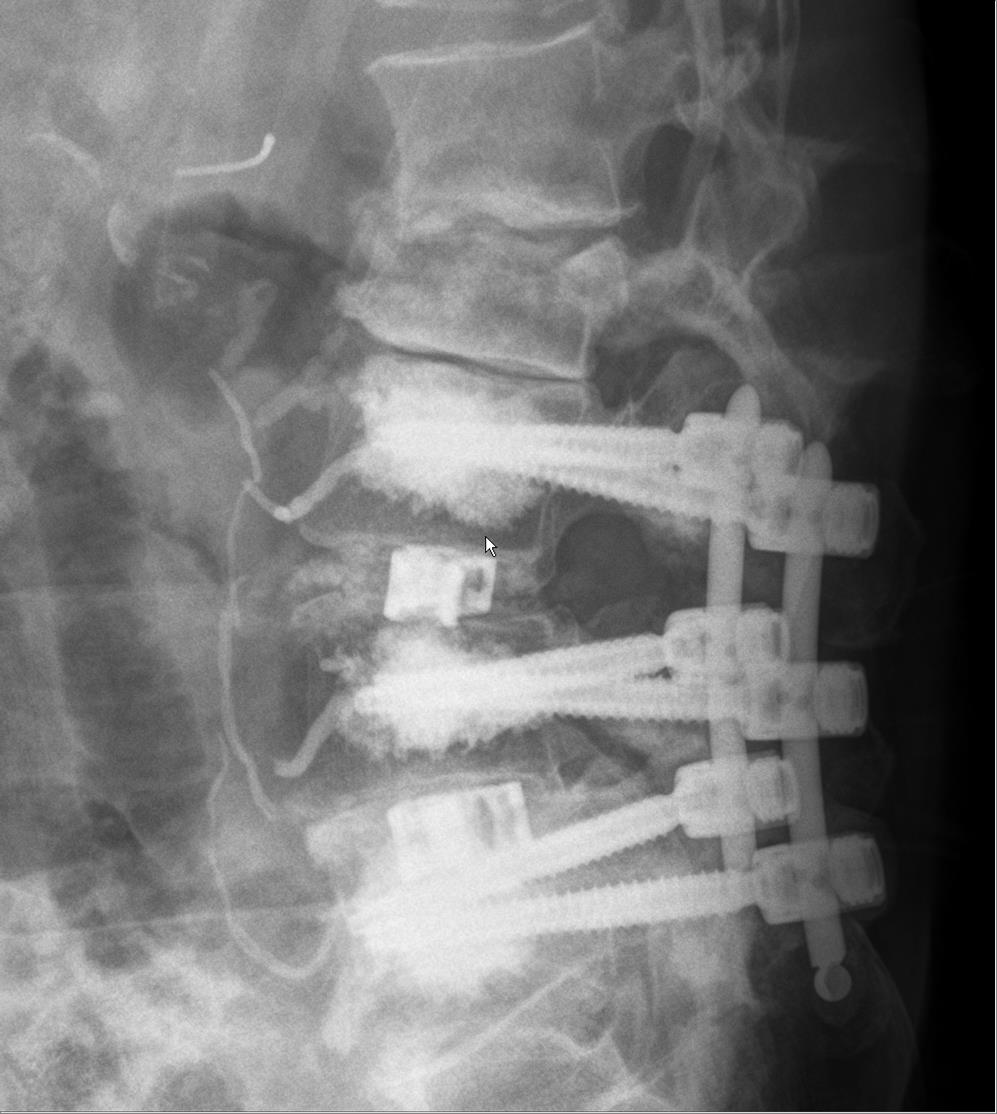
Fixation in osteoporotic bone
- Expect screw pull-out and loosening; plan for it rather than discover it
- Cement-augmented pedicle screws: pooled loosening 2.0%, breakage 0.6%; any cement leakage 21.8% but symptomatic leakage 1.2%, symptomatic pulmonary embolism 0.8% Zhang 2021
- PMMA and calcium-based cements both increase pull-out strength; fenestrated screws and pre-fill techniques perform similarly Elder 2015
- Longer constructs, more fixation points, and hooks or bands to share load; avoid ending a construct at the apex of a kyphosis or next to a fractured level
- Reconstruct the anterior column when it cannot bear load: augmentation of the index vertebra, cage, or strut; PMMA, not calcium phosphate, for stand-alone fill in osteoporotic bone Uchida 2010 Sudo 2013 Schmidt 2011
- Restore the patient's own pre-fracture sagittal profile, not a textbook one; minimise invasiveness in this population Blattert 2018
- Treat the bone: bone-health optimisation improves outcomes and anabolic therapy is favoured around instrumentation Anderson 2019
Treating the bone
The strongest evidence in the pathway and the worst compliance: the drugs by name, what they do, and in what order
We know what to do and we do not do it
The bone-health work-up
- A vertebral fragility fracture after 50 is diagnostic of osteoporosis regardless of the T-score, and signals imminent risk of the next fracture LeBoff 2022
- DXA of hip and spine to stage severity and to monitor; screen women ≥ 65 and younger postmenopausal women at increased risk US 2025
- FRAX 10-year probability, with or without BMD, adjusted for the recent fracture Kanis 2008
- Laboratory screen for secondary causes: calcium, phosphate, alkaline phosphatase, renal function, 25-OH vitamin D, TSH, full blood count, protein electrophoresis; testosterone in men; consider PTH and coeliac serology
- Opportunistic CT: lumbar Hounsfield units on any CT already performed correlate with DXA and strength Schreiber 2011 Anderson 2019
- Orthopaedic surgeons should own this: DXA before surgery in high-risk patients, correct deficits, and consider delaying elective surgery 3 months to start treatment Anderson 2019 Anderson 2019
What each antiresorptive does to fracture risk
| Agent (US brand), class, dose | Pivotal trial | New vertebral fractures | Hip / nonvertebral | Watch for |
|---|---|---|---|---|
| Alendronate (Fosamax), bisphosphonate, 70 mg PO weekly | FIT, 2,027 women with prior VF, 3 y Black 1996 | 8.0% vs 15.0%, RR 0.53 (−47%) | Hip RH 0.49; wrist RH 0.52 | GI intolerance; AFF and ONJ with long use |
| Risedronate (Actonel; Atelvia delayed-release), bisphosphonate, 35 mg PO weekly | VERT-NA, 2,458 women, 3 y Harris 1999 | 11.3% vs 16.3% (−41%); −65% in year 1 | Nonvertebral 5.2% vs 8.4% (−39%) | As alendronate |
| Ibandronate (Boniva), bisphosphonate, 150 mg PO monthly | BONE, 2,946 women, 3 y Chesnut 2004 | 4.7% vs 9.6% (−62%) | No hip or nonvertebral benefit | Spine-only agent |
| Zoledronic acid (Reclast), bisphosphonate, 5 mg IV yearly | HORIZON, 7,765 women, 3 y Black 2007 | 3.3% vs 10.9%, RR 0.30 (−70%) | Hip 1.4% vs 2.5% (−41%); nonvertebral −25% | Acute-phase reaction; eGFR ≥ 35; AF signal |
| Denosumab (Prolia), RANKL antibody, 60 mg SC 6-monthly | FREEDOM, 7,868 women, 3 y Cummings 2009 | 2.3% vs 7.2%, RR 0.32 (−68%) | Hip −40%; nonvertebral −20% | Rebound fractures if stopped without a bisphosphonate |
| Raloxifene (Evista), SERM, 60 mg PO daily | MORE, 7,705 women, 3 y Ettinger 1999 | 6.6% vs 10.1%, RR 0.7 (−30%) | None | VTE RR 3.1; spine-only |
New morphometric vertebral fractures, drug versus placebo. Replete vitamin D and check renal function before any bisphosphonate.
Build bone first in the patient who has just fractured
| Agent (US brand), class, dose | Pivotal trial | New vertebral fractures | Hip / nonvertebral | Watch for |
|---|---|---|---|---|
| Teriparatide (Forteo; Bonsity), PTH 1–34, 20 µg SC daily, up to 2 y | Neer 2001, 1,637 women with prior VF, 21 mo Neer 2001 | 5% vs 14%, RR 0.35 (−65%) | Nonvertebral fragility 3% vs 6% (−53%) | Nausea, cramps, hypercalcaemia; not in Paget disease, open physes or prior skeletal radiation |
| Teriparatide vs risedronate | VERO, 1,360 women with severe osteoporosis, 2 y Kendler 2018 | 5.4% vs 12.0%, RR 0.44 | Clinical fractures HR 0.48 | Head-to-head win for the anabolic |
| Abaloparatide (Tymlos), PTHrP analogue, 80 µg SC daily, up to 2 y | ACTIVE, 2,463 women, 18 mo Miller 2016 | 0.58% vs 4.22% (−86%) | Nonvertebral HR 0.57 (−43%) | Less hypercalcaemia than teriparatide (3.4% vs 6.4%) |
| Romosozumab (Evenity), anti-sclerostin, 210 mg SC monthly × 12 | FRAME, 7,180 women, 12 mo Cosman 2016 | 0.5% vs 1.8% (−73%) | Clinical fractures −36%; nonvertebral NS at 12 mo | Then denosumab: vertebral fractures −75% at 24 mo |
| Romosozumab → alendronate | ARCH, 4,093 women with a fragility fracture, 2 y Saag 2017 | 6.2% vs 11.9% (−48%) vs alendronate alone | Hip −38%; nonvertebral −19% | Serious cardiovascular events 2.5% vs 1.9% in year 1: boxed warning Shoback 2020 |
Network meta-analysis of 69 trials: anabolics beat oral bisphosphonates for vertebral and clinical fractures at every level of baseline risk. Händel 2023
Sequence, duration and the safety conversation
- Anabolic first, antiresorptive second: teriparatide then denosumab gained 18% spine and 7% hip BMD over 4 years; the reverse order lost hip and radius bone during the switch Leder 2015
- Every anabolic course ends with an antiresorptive or the gain is lost. One year of Evenity then Prolia reaches a T-score that takes about 7 years of Prolia alone Cosman 2018; after years of a bisphosphonate, Evenity still adds hip BMD where Forteo first loses it Langdahl 2017
- Prolia is a commitment: stopping without a bisphosphonate raises the vertebral fracture rate from 1.2 to 7.1 per 100 patient-years, 61% of them multiple Cummings 2018; 10 years of continuous use stays safe and effective Bone 2017
- Bisphosphonate holiday after 5 years oral or 3 years IV is for the patient without a vertebral fracture: in FLEX, stopping doubled clinical vertebral fractures (5.3% vs 2.4%) while nonvertebral risk was unchanged Black 2006
- Atypical femoral fracture: HR 8.9 at 3–5 years and 43.5 at ≥ 8 years of bisphosphonate, falling fast after stopping; 149 hip fractures prevented per 2 atypical fractures Black 2020. Jaw osteonecrosis 0.001–0.01% at osteoporosis doses Khan 2015
Which drug, and when, after a vertebral fracture
- A recent vertebral fracture places the patient at very high risk: anabolic-first sequences are endorsed for this group Camacho 2020 Qaseem 2023
- In patients with prevalent vertebral fractures, anabolics halve new vertebral fractures compared with bisphosphonates (RR 0.57, high certainty) Jeon 2024
- Fracture healing: teriparatide raised 6-month union from 68% to 89% versus alendronate in one series, but pooled RCT evidence for healing is low-certainty and negative Iwata 2017 Jeon 2024; bisphosphonates do not impair vertebral healing and can start as soon as the pain allows
- Calcium and vitamin D repletion, falls assessment, smoking and alcohol counselling in every patient LeBoff 2022
- Around fusion: daily teriparatide raised posterolateral fusion from 68% to 82% versus risedronate and shortened union by 2 months Ohtori 2012; a randomised trial of weekly teriparatide after PLIF/TLIF showed higher CT fusion at 4 and 6 months Ebata 2017
Two cases
Apply it
The 78-year-old from the first slide
- Decision and course
- Discussed: sham trials show small average benefit; she fits the subgroup (acute, edema, severe pain, functional failure) where trials and pathways support offering it
- L1 balloon kyphoplasty at 4 weeks; walking the same evening, pain 3/10 at 2 weeks
- DXA T-score −3.4 spine; started teriparatide with calcium and vitamin D, fracture liaison follow-up; no new fracture at 18 months
An 82-year-old man with a burst fracture
- Course
- Mobilised day 2 with physiotherapy; pneumonia day 4, treated; discharged to rehabilitation day 12
- At 3 months: walking with a frame, dorsiflexion 4/5, voiding spontaneously
- DXA T-score −3.1; started zoledronate after vitamin D repletion (anabolic declined: cost and injections); fracture liaison follow-up
A 74-year-old woman is diagnosed with an acute osteoporotic L2 compression fracture and treated nonoperatively. Her DXA T-score is −2.9 and this is her first fracture. Which statement about secondary prevention is best supported by evidence?
- AWait 6 months for the fracture to heal before starting any osteoporosis medication, because bisphosphonates impair healing
- BA single vertebral fracture does not change management; repeat DXA in 2 years
- CShe is at very high risk of another fracture within a year; start treatment now, with an anabolic-first sequence endorsed for very-high-risk patients and a bisphosphonate as the standard first-line alternative
- DVertebral augmentation is the most effective way to prevent her next fracture
Seven things to take back to the department
- A vertebral fragility fracture is a sentinel event: 1 in 5 refracture within a year and mortality is doubled. Look for it on every chest and abdominal film
- Standing radiographs first; MRI with STIR when the age is uncertain, there are red flags or a deficit, or a treatment decision depends on it; CT for the posterior wall
- Convex posterior cortex, pedicle signal, or a soft-tissue mass means malignancy until proven otherwise
- Classify: Genant for severity, TLICS to communicate, AOSpine with the M2 modifier to describe, OF type to plan. Posterior wall, cleft, and neurology are the pivots
- Nonoperative care is a package: analgesia, same-day mobilisation, safe movement, standing films at 2–4, 6 and 12 weeks. A rigid brace does not prevent collapse
- Augmentation is not routine; it is a reasonable option for MRI-positive, severe, refractory, function-limiting pain after an honest discussion of the sham trials
- Treat the bone before the patient leaves: labs, DXA, FRAX, an anabolic first (Forteo, Tymlos or Evenity) then an antiresorptive, never Prolia without an exit plan, and a fracture liaison service
The fracture in front of you is the best predictor of the fracture you can still prevent.
Find it, classify it, treat the pain honestly, refer the ones that are not benign, and treat the bone.
Selected references
1.Lindsay R, Silverman SL, Cooper C, Hanley DA, Barton I, Broy SB. Risk of new vertebral fracture in the year following a fracture. JAMA. 2001;285(3):320-3.
2.Kado DM, Browner WS, Palermo L, Nevitt MC, Genant HK, Cummings SR. Vertebral fractures and mortality in older women: a prospective study. Study of Osteoporotic Fractures Research Group. Arch Intern Med. 1999;159(11):1215-20.
3.Lau E, Ong K, Kurtz S, Schmier J, Edidin A. Mortality following the diagnosis of a vertebral compression fracture in the Medicare population. J Bone Joint Surg Am. 2008;90(7):1479-86.
4.Delmas PD, van de Langerijt L, Watts NB, Eastell R, Genant H, Grauer A. Underdiagnosis of vertebral fractures is a worldwide problem: the IMPACT study. J Bone Miner Res. 2005;20(4):557-63.
5.Genant HK, Wu CY, van Kuijk C, Nevitt MC. Vertebral fracture assessment using a semiquantitative technique. J Bone Miner Res. 1993;8(9):1137-48.
6.Schnake KJ, Blattert TR, Hahn P, Franck A, Hartmann F, Ullrich B. Classification of Osteoporotic Thoracolumbar Spine Fractures: Recommendations of the Spine Section of the German Society for Orthopaedics and Trauma (DGOU). Global Spine J. 2018;8(2 Suppl):46S-49S.
7.Blattert TR, Schnake KJ, Gonschorek O, Gercek E, Hartmann F, Katscher S. Nonsurgical and Surgical Management of Osteoporotic Vertebral Body Fractures: Recommendations of the Spine Section of the German Society for Orthopaedics and Trauma (DGOU). Global Spine J. 2018;8(2 Suppl):50S-55S.
8.Vaccaro AR, Oner C, Kepler CK, Dvorak M, Schnake K, Bellabarba C. AOSpine thoracolumbar spine injury classification system: fracture description, neurological status, and key modifiers. Spine (Phila Pa 1976). 2013;38(23):2028-37.
9.Jung HS, Jee WH, McCauley TR, Ha KY, Choi KH. Discrimination of metastatic from acute osteoporotic compression spinal fractures with MR imaging. Radiographics. 2003;23(1):179-87.
10.Tsujio T, Nakamura H, Terai H, Hoshino M, Namikawa T, Matsumura A. Characteristic radiographic or magnetic resonance images of fresh osteoporotic vertebral fractures predicting potential risk for nonunion: a prospective multicenter study. Spine (Phila Pa 1976). 2011;36(15):1229-35.
11.Muratore M, Ferrera A, Masse A, Bistolfi A. Osteoporotic vertebral fractures: predictive factors for conservative treatment failure. A systematic review. Eur Spine J. 2018;27(10):2565-2576.
12.Kim HJ, Yi JM, Cho HG, Chang BS, Lee CK, Kim JH. Comparative study of the treatment outcomes of osteoporotic compression fractures without neurologic injury using a rigid brace, a soft brace, and no brace: a prospective randomized controlled non-inferiority trial. J Bone Joint Surg Am. 2014;96(23):1959-66.
13.Kato T, Inose H, Ichimura S, Tokuhashi Y, Nakamura H, Hoshino M. Comparison of Rigid and Soft-Brace Treatments for Acute Osteoporotic Vertebral Compression Fracture: A Prospective, Randomized, Multicenter Study. J Clin Med. 2019;8(2).
14.Buchbinder R, Osborne RH, Ebeling PR, Wark JD, Mitchell P, Wriedt C. A randomized trial of vertebroplasty for painful osteoporotic vertebral fractures. N Engl J Med. 2009;361(6):557-68.
15.Kallmes DF, Comstock BA, Heagerty PJ, Turner JA, Wilson DJ, Diamond TH. A randomized trial of vertebroplasty for osteoporotic spinal fractures. N Engl J Med. 2009;361(6):569-79.
16.Klazen CA, Lohle PN, de Vries J, Jansen FH, Tielbeek AV, Blonk MC. Vertebroplasty versus conservative treatment in acute osteoporotic vertebral compression fractures (Vertos II): an open-label randomised trial. Lancet. 2010;376(9746):1085-92.
17.Firanescu CE, de Vries J, Lodder P, Venmans A, Schoemaker MC, Smeets AJ. Vertebroplasty versus sham procedure for painful acute osteoporotic vertebral compression fractures (VERTOS IV): randomised sham controlled clinical trial. BMJ. 2018;361:k1551.
18.Clark W, Bird P, Gonski P, Diamond TH, Smerdely P, McNeil HP. Safety and efficacy of vertebroplasty for acute painful osteoporotic fractures (VAPOUR): a multicentre, randomised, double-blind, placebo-controlled trial. Lancet. 2016;388(10052):1408-1416.
19.Wardlaw D, Cummings SR, Van Meirhaeghe J, Bastian L, Tillman JB, Ranstam J. Efficacy and safety of balloon kyphoplasty compared with non-surgical care for vertebral compression fracture (FREE): a randomised controlled trial. Lancet. 2009;373(9668):1016-24.
20.Buchbinder R, Johnston RV, Rischin KJ, Homik J, Jones CA, Golmohammadi K. Percutaneous vertebroplasty for osteoporotic vertebral compression fracture. Cochrane Database Syst Rev. 2018;11(11):CD006349.
21.Ebeling PR, Akesson K, Bauer DC, Buchbinder R, Eastell R, Fink HA. The Efficacy and Safety of Vertebral Augmentation: A Second ASBMR Task Force Report. J Bone Miner Res. 2019;34(1):3-21.
22.Hirsch JA, Beall DP, Chambers MR, Andreshak TG, Brook AL, Bruel BM. Management of vertebral fragility fractures: a clinical care pathway developed by a multispecialty panel using the RAND/UCLA Appropriateness Method. Spine J. 2018;18(11):2152-2161.
23.Carli D, Venmans A, Lodder P, Donga E, van Oudheusden T, Boukrab I. Vertebroplasty versus Active Control Intervention for Chronic Osteoporotic Vertebral Compression Fractures: The VERTOS V Randomized Controlled Trial. Radiology. 2023;308(1):e222535.
24.Uchida K, Nakajima H, Yayama T, Miyazaki T, Hirai T, Kobayashi S. Vertebroplasty-augmented short-segment posterior fixation of osteoporotic vertebral collapse with neurological deficit in the thoracolumbar spine: comparisons with posterior surgery without vertebroplasty and anterior surgery. J Neurosurg Spine. 2010;13(5):612-21.
25.Black DM, Cummings SR, Karpf DB, Cauley JA, Thompson DE, Nevitt MC. Randomised trial of effect of alendronate on risk of fracture in women with existing vertebral fractures. Fracture Intervention Trial Research Group. Lancet. 1996;348(9041):1535-41.
26.Saag KG, Petersen J, Brandi ML, Karaplis AC, Lorentzon M, Thomas T. Romosozumab or Alendronate for Fracture Prevention in Women with Osteoporosis. N Engl J Med. 2017;377(15):1417-1427.
27.Kendler DL, Marin F, Zerbini CAF, Russo LA, Greenspan SL, Zikan V. Effects of teriparatide and risedronate on new fractures in post-menopausal women with severe osteoporosis (VERO): a multicentre, double-blind, double-dummy, randomised controlled trial. Lancet. 2018;391(10117):230-240.
28.Anderson PA, Jeray KJ, Lane JM, Binkley NC. Bone Health Optimization: Beyond Own the Bone: AOA Critical Issues. J Bone Joint Surg Am. 2019;101(15):1413-1419.
29.Wu CH, Tu ST, Chang YF, Chan DC, Chien JT, Lin CH. Fracture liaison services improve outcomes of patients with osteoporosis-related fractures: A systematic literature review and meta-analysis. Bone. 2018;111:92-100.
30.Cho CH, Hwang SW, Mazanec DJ, O'Toole JE, Watters WC 3rd, Annaswamy TM. Guideline summary review: an evidence-based clinical guideline for the diagnosis and treatment of adults with osteoporotic vertebral compression fractures. Spine J. 2025;25(8):1670-1687.
31.Expert Panels on Neurological Imaging, Interventional Radiology, and Musculoskeletal Imaging, Khan MA, Jennings JW, Baker JC, Smolock AR, Shah LM. ACR Appropriateness Criteria® Management of Vertebral Compression Fractures: 2022 Update. J Am Coll Radiol. 2023;20(5S):S102-S124.
32.Qaseem A, Hicks LA, Etxeandia-Ikobaltzeta I, Shamliyan T, Cooney TG, Clinical Guidelines Committee of the American College of Physicians. Pharmacologic Treatment of Primary Osteoporosis or Low Bone Mass to Prevent Fractures in Adults: A Living Clinical Guideline From the American College of Physicians. Ann Intern Med. 2023;176(2):224-238.
33.Pieroh P, Spiegl UJA, Völker A, Märdian S, von der Höh NH, Osterhoff G. Spinal Orthoses in the Treatment of Osteoporotic Thoracolumbar Vertebral Fractures in the Elderly: A Systematic Review With Quantitative Quality Assessment. Global Spine J. 2023;13(1_suppl):59S-72S.
34.Zhang J, Wang G, Zhang N. A meta-analysis of complications associated with the use of cement-augmented pedicle screws in osteoporosis of spine. Orthop Traumatol Surg Res. 2021;107(7):102791.
35.Wiedl A, Geiger F, Eberl J, Wirries A, Hammad A. Mortality After Osteoporotic Vertebral Body Fractures: Cement Augmentation Versus Conservative Treatment—A Systematic Review and Meta-Analysis. Dtsch Arztebl Int. 2025;122(22):604-610.
36.Denis F. The three column spine and its significance in the classification of acute thoracolumbar spinal injuries. Spine (Phila Pa 1976). 1983;8(8):817-31.
37.Vaccaro AR, Lehman RA Jr, Hurlbert RJ, Anderson PA, Harris M, Hedlund R. A new classification of thoracolumbar injuries: the importance of injury morphology, the integrity of the posterior ligamentous complex, and neurologic status. Spine (Phila Pa 1976). 2005;30(20):2325-33.
38.Kepler CK, Vaccaro AR, Schroeder GD, Koerner JD, Vialle LR, Aarabi B. The Thoracolumbar AOSpine Injury Score. Global Spine J. 2016;6(4):329-34.
39.Black DM, Delmas PD, Eastell R. Once-yearly zoledronic acid for treatment of postmenopausal osteoporosis. N Engl J Med. 2007;356(18):1809-22.
40.Cosman F, Crittenden DB, Adachi JD. Romosozumab Treatment in Postmenopausal Women with Osteoporosis. N Engl J Med. 2016;375(16):1532-1543.
41.Wood K, Buttermann G, Mehbod A, Garvey T, Jhanjee R, Sechriest V. Operative compared with nonoperative treatment of a thoracolumbar burst fracture without neurological deficit. A prospective, randomized study. J Bone Joint Surg Am. 2003;85(5):773-81.
42.Bailey CS, Urquhart JC, Dvorak MF, Nadeau M, Boyd MC, Thomas KC. Orthosis versus no orthosis for the treatment of thoracolumbar burst fractures without neurologic injury: a multicenter prospective randomized equivalence trial. Spine J. 2014;14(11):2557-64.
43.Tezeren G, Kuru I. Posterior fixation of thoracolumbar burst fracture: short-segment pedicle fixation versus long-segment instrumentation. J Spinal Disord Tech. 2005;18(6):485-8.
Full reference list with DOI and PubMed links is available in the online references view.
Slides, notes and references
Scan for the full reference list with PubMed links, the speaker notes handout, and this deck.