Osteoporotic Vertebral Fractures
Speaker notes · 50 min · v1.2 · updated 2026-09-27
- 1 / 60~1 min · at 0:30Introduction
Welcome. This talk is about the most common osteoporotic fracture, the one most often missed, and the one that most reliably predicts the next fracture: the vertebral compression fracture.
Two promises. First, by the end you will be able to sort an older patient with acute back pain into "stable fracture, treat the pain and the bone," "fracture that needs a spine surgeon," or "this is not osteoporosis." Second, I will give you the honest state of the evidence on vertebroplasty and kyphoplasty, which is more contested than for almost any other procedure we perform, and tell you what the guidelines actually say.
Every claim is referenced; the chips on the slides open the source, and the full list with PubMed links is online.
- 2 / 60~1 min · at 1:15Introduction
Start with the patient because the failure mode is so common it has a name in the literature: underdiagnosis and undertreatment.
Paint the picture. A low-energy event, sometimes no event at all. Acute midline back pain in a woman in her late seventies. A radiograph that shows a wedge at L1 and a report that hedges on its age. She goes home with an opioid prescription and a follow-up with her primary care physician that may or may not happen.
What did not happen: nobody established whether the fracture was acute, nobody asked whether the posterior wall was intact, nobody assessed her bone health, and nobody started treatment that would have cut her risk of the next fracture by half. Eleven months later she has a second fracture, and the literature says that is exactly what to expect: one in five patients with a new vertebral fracture has another within a year Lindsay 2001.
Keep her in mind; she returns in the cases.
Pearl: The first vertebral fracture is a diagnostic opportunity for the skeleton, not just a back pain problem. - 3 / 60~1 min · at 1:45Introduction
Seven objectives, deliberately spanning the whole pathway from the emergency department to the osteoporosis clinic, because the biggest gains for this patient population are at the two ends: recognising the fracture, and treating the bone.
Objective five is where I will spend the most time on evidence, because the trials disagree with each other and the guidelines disagree with the trials.
- 4 / 60~0 min · at 1:55Why it matters
Section one, about four minutes: the scale of the problem and the consequences of missing it.
- 5 / 60~1 min · at 3:10Why it matters
Four numbers that frame the problem.
Worldwide, in the year 2000, there were about nine million osteoporotic fractures, of which 1.4 million were clinical vertebral fractures, and the disability burden exceeded that of most cancers in Europe Johnell 2006. Clinically diagnosed vertebral fractures occur at roughly 117 per 100,000 person-years in the Rochester population, twice as often in women as men, and 83% follow moderate or no trauma Cooper 1992. Remember that clinically diagnosed fractures are only about a third of all vertebral fractures; the rest are silent or misattributed.
The second half of the slide is about missing them. In the IMPACT study, 2,451 osteoporotic women had spine radiographs read locally and then centrally: the local false-negative rate was 34%, and in North America 45% Delmas 2005. Gehlbach looked at 934 hospitalised older women who happened to have chest radiographs: 14% had a moderate or severe vertebral fracture visible on the film, 1.8% had it recorded as a diagnosis, and 18% received any osteoporosis treatment Gehlbach 2000.
So the fracture is common, it is on the films we already take, and we do not see it or act on it. Two habits fix most of the underdiagnosis: look at the spine on every lateral chest and abdominal film, and use the word "fracture" in the report rather than "deformity" or "loss of height," because equivocal wording accounted for a third of the missed diagnoses in IMPACT Delmas 2005.
- 6 / 60~1 min · at 3:55Why it matters
This is the single most important number in the talk.
Lindsay analysed the placebo arms of four large osteoporosis trials, 2,725 women with a mean age of 74. Among the 381 who sustained an incident vertebral fracture during the study, 19.2% had another vertebral fracture within the following year. Having a prevalent fracture at baseline raised the one-year risk five-fold Lindsay 2001. Klotzbuecher's synthesis put the long-term risk at about four-fold for a further vertebral fracture and two-fold for fractures elsewhere Klotzbuecher 2000.
This is why the fracture is a sentinel event: the period of highest risk is the year immediately after, which is exactly when we see the patient. Anti-osteoporotic medication reduces subsequent vertebral fractures by 40 to 70% Ebeling 2019. Not treating the bone after a vertebral fracture is a missed opportunity measured in fractures.
Refs: Lindsay et al., JAMA, 2001 · Klotzbuecher et al., J Bone Miner Res, 2000 - 7 / 60~1 min · at 5:10Why it matters
Vertebral fractures shorten life, and the mechanism is mostly respiratory.
In the Study of Osteoporotic Fractures, 9,575 women followed for eight years, a prevalent vertebral fracture was associated with a 1.23-fold higher age-adjusted mortality, rising with the number of fractures from 19 to 44 deaths per 1,000 woman-years, with a doubling of pulmonary death and a 2.6-fold pulmonary mortality in those with severe kyphosis Kado 1999. In the FIT cohort, prevalent deformities predicted both mortality and hospitalisation Ensrud 2000.
In Medicare data, 97,142 patients with a newly diagnosed compression fracture had roughly twice the mortality of matched controls, with survival of 54%, 31% and 10% at three, five and seven years Lau 2008. The mortality gap was larger for men and for younger patients. Lau was careful to say this does not prove causation; the fracture marks a frail skeleton and a frail patient.
For the inpatients we admit, the systematic review by Ong found a mean age of 81, a median stay of ten days, one-year mortality of 20 to 27%, and a third to a half discharged to care facilities, with only about a third started on osteoporosis treatment Ong 2018.
The physiological link is the chest: thoracic kyphosis and loss of height reduce vital capacity, about 12 percentage points of predicted in Schlaich's series, in proportion to the spinal deformity Schlaich 1998. Each new fracture makes breathing a little harder.
- 8 / 60~0 min · at 5:20Mechanism & classification
Section two, about seven minutes: the mechanics of the osteoporotic vertebra and the classifications you will hear surgeons use.
Why the vertebra fails
- Osteoporosis thins and disconnects the trabecular network; the vertebral body is mostly trabecular bone and fails under axial load and flexion
- Thoracolumbar junction is the commonest site: L1 in 19% of one inpatient series, with T12 and L2 close behind Schnake 2022
- Minimal or no trauma: 83% of clinical fractures follow moderate or no trauma; half of hospitalised patients recall no injury Cooper 1992 Schnake 2022
- Anterior wedge is typical; end-plate failure gives biconcave and crush patterns
- Some fractures keep moving: 44% of vertebroplasty candidates showed dynamic mobility between standing and supine films McKiernan 2003
- Progression of deformity is linked to a T-score below −3 Schnake 2022
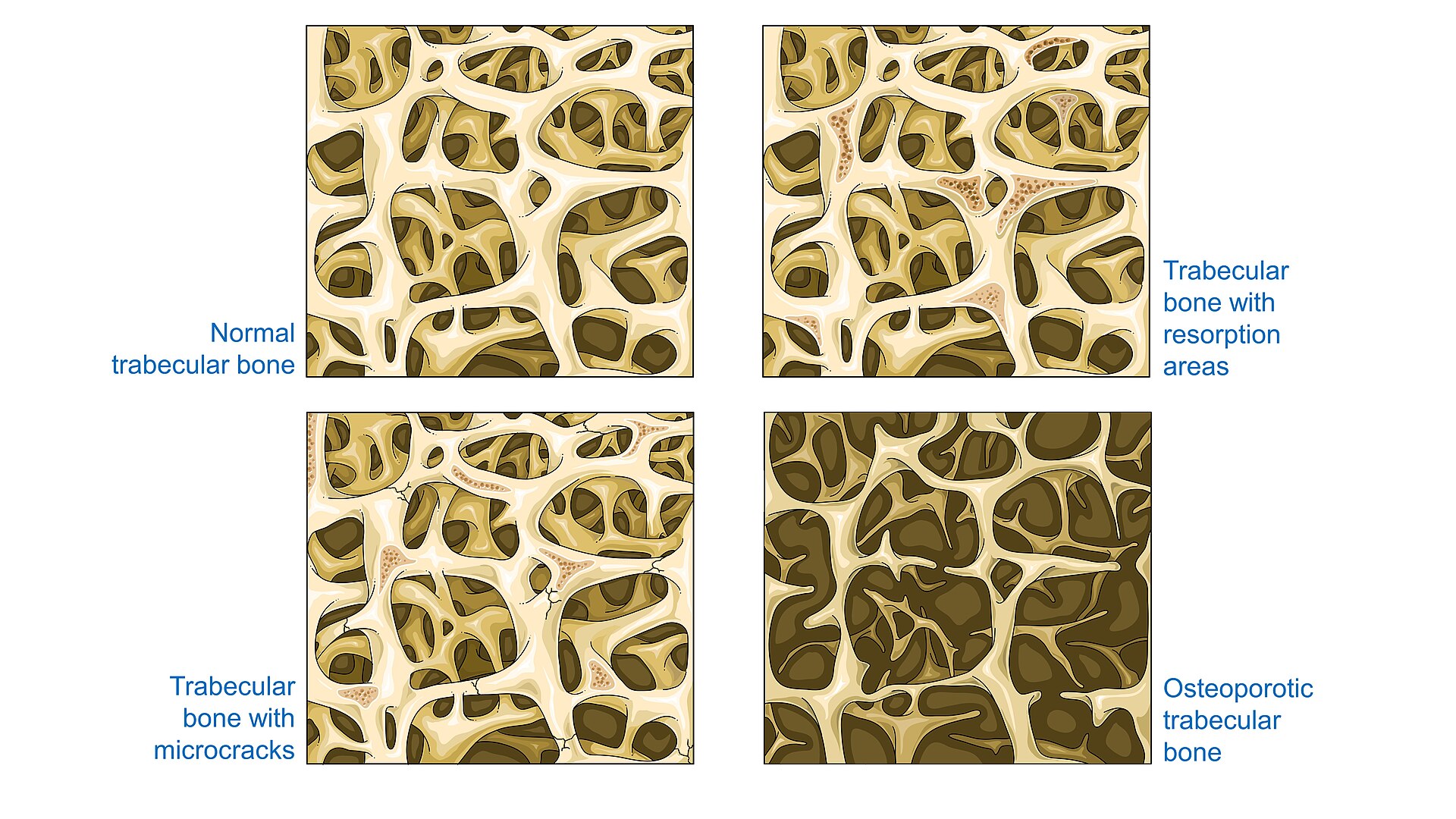
Normal trabecular bone, resorption, microcracks, osteoporotic bone. 9 / 60~1 min · at 6:35Mechanism & classificationThe vertebral body is a shell of thin cortical bone around a block of trabecular bone, and osteoporosis attacks the trabeculae first, thinning the horizontal struts and disconnecting the network. The result is a structure that fails under ordinary axial loads and flexion moments: lifting, coughing, sitting down hard, or nothing at all.
The thoracolumbar junction takes the brunt because it is where the stiff kyphotic thoracic spine meets the mobile lumbar spine. In the German multicentre inpatient study of 707 patients, L1 alone accounted for 19% of fractures, more than half the patients could not remember a traumatic event, and progression of deformity was associated with a T-score below minus three Schnake 2022. In the Rochester data 83% of clinical fractures followed moderate or no trauma Cooper 1992.
The classic pattern is the anterior wedge. End-plate failure gives the biconcave "codfish" vertebra, and a crush fracture loses height throughout.
One detail that matters for imaging and for augmentation: McKiernan showed that 44% of patients referred for vertebroplasty had fractures that changed shape between standing and supine radiographs, and every mobile fracture had an intravertebral cleft McKiernan 2003. That mobility is why standing films matter, why some "height restoration" after augmentation is simply positioning, and why a cleft is a warning sign we will return to.
Refs: Schnake et al., Eur J Trauma Emerg Surg, 2022 · Cooper et al., J Bone Miner Res, 1992 · McKiernan et al., J Bone Miner Res, 2003- 10 / 60~1 min · at 7:35Mechanism & classification
Genant's semiquantitative grading is the language of the osteoporosis literature and of every drug trial Genant 1993. It is worth knowing because it also defines who has "severe" osteoporosis in the treatment guidelines.
Morphology first: wedge, with anterior height loss; biconcave, with central end-plate depression; and crush, with loss of all heights. Then severity: grade 1 is 20 to 25% height loss compared with the vertebra's expected height, grade 2 is 25 to 40%, and grade 3 is more than 40%.
Two practical points. A 20% threshold means that many real fractures are subtle on a single film and are easily called "degenerative" or "age indeterminate"; comparison with prior films is invaluable. And the drug trials of anabolic agents enrolled women with at least two moderate or one severe fracture, so a grade 3 fracture on your film is, in itself, an indication to think about anabolic therapy Kendler 2018.
Refs: Genant et al., J Bone Miner Res, 1993 - 11 / 60~1 min · at 9:00Mechanism & classification
Trauma classifications like AOSpine were built for young bone and instability Vaccaro 2013. The German Society for Orthopaedics and Trauma built a classification specifically for osteoporotic fractures, now adopted by AO Spine, that describes what actually happens to these vertebrae Schnake 2018.
Walk through the five types. OF1: no deformation, only bone-marrow edema on MRI; the painful vertebra that looks normal on the radiograph. OF2: deformation of the anterior wall only, with no or minimal, less than one fifth, posterior wall involvement; the classic wedge. OF3: distinct posterior wall involvement, more than one fifth, the incomplete burst. OF4: loss of integrity of the vertebral frame, vertebral body collapse, or a pincer fracture; this includes the burst-split and the Kümmell collapse. OF5: injuries with distraction or rotation, which are the unstable ligamentous injuries, often in ankylosed spines.
Reliability: substantial in the original description, kappa 0.63 Schnake 2018, and moderate, around 0.5, with substantial intra-rater reproducibility in the AO Spine international validation Scherer 2025. Like every classification, it improves with training.
The reason to learn it is that the DGOU treatment recommendations hang on it: an OF score below 6 favours nonoperative care, above 6 favours surgery, and the type is upgraded on follow-up films if the fracture progresses Blattert 2018. OF1 and OF2 are usually nonoperative; OF4 and OF5 are usually surgical; OF3 is the individualised middle.
Refs: Schnake et al., Global Spine J, 2018 · Scherer et al., Global Spine J, 2025 - 12 / 60~1 min · at 10:00Mechanism & classification
Residents will be asked about all of these, so here is the lineage in one line.
Denis analysed 412 CT-imaged injuries in 1983 and gave us the three-column spine: anterior column of the anterior longitudinal ligament and anterior body, middle column of the posterior body and posterior longitudinal ligament, posterior column of the arch, facets and ligaments. Compression fractures fail the anterior column; burst fractures fail the anterior and middle columns; instability begins when two columns fail Denis 1983. It is still the vocabulary we use at the bedside.
Magerl's AO classification of 1994, from 1,445 injuries, organised fractures by mechanism into type A compression, B distraction and C rotation, with 27 subgroups; type A1 impaction fractures alone were a third of all injuries Magerl 1994 McCall 2011. Comprehensive, but in reliability studies the agreement was only fair to moderate and fell further with every subgroup Oner 2002 Wood 2005.
In 2005 the Spine Trauma Study Group replaced mechanism with morphology, added the posterior ligamentous complex and the neurological examination, and attached points that recommend treatment: the Thoracolumbar Injury Classification and Severity Score Vaccaro 2005. The next two slides go through it.
In 2013 AOSpine reconciled the two traditions in an international consensus: A0 to A4, B1 to B3 and C for morphology, a neurological grade and patient-specific modifiers, with substantial reliability for the type Vaccaro 2013.
And in 2018 the German trauma society built the OF classification specifically for the low-energy osteoporotic fracture, because none of the trauma systems described what happens to these vertebrae over the following weeks Schnake 2018. That is where we ended up on the previous slide; now the trauma systems in detail.
- 13 / 60~2 min · at 11:40Mechanism & classification
TLICS in four steps.
Step one, morphology. Compression scores 1, burst 2, translation or rotation 3, distraction 4. In a combined pattern only the most severe descriptor is scored. Burst fractures automatically score 2, which is why an isolated osteoporotic burst without PLC injury or deficit lands at 2 and nonoperative care Vaccaro 2005 Petfield 2011.
Step two, the posterior ligamentous complex: supraspinous and interspinous ligaments, ligamentum flavum and facet capsules, which resist flexion, distraction, rotation and translation and heal poorly in adults. Intact 0, indeterminate 2, disrupted 3. Clinically, palpate for a gap between spinous processes; on imaging look for splaying of the spinous processes, facet subluxation or perching, vertebral translation, and edema through the PLC on STIR or fat-suppressed T2, which the Spine Trauma Study Group specifically recommended to improve reliability Vaccaro 2006.
Step three, neurology, from the ISNCSCI examination: intact 0, nerve root 2, complete cord or conus 2, incomplete cord or conus 3, cauda equina 3. Incomplete injuries score highest because they have the most to gain from decompression.
Step four, add them. Three or less: nonoperative. Five or more: operative. Four: the grey zone, decided by the surgeon and the patient's circumstances.
The validation data: in the original reliability study five surgeons agreed with the score's recommendation in more than 90% of 71 cases Vaccaro 2006; Whang found TLISS and TLICS both valid, with TLICS predicting treatment in 95% Whang 2007; Patel showed reliability improves with use, so it can be taught Patel 2007; and Joaquim found the TLICS recommendation matched the surgery actually performed in 96% of 49 operated patients Joaquim 2011. Bono's caveat still stands: the third phase of validation, prospective outcome studies, was never completed in the way Audigé's framework demands Bono 2006.
TLICS was also built to suggest the approach. Anterior compression of the cord with an incomplete deficit argued for anterior decompression; a disrupted posterior ligamentous complex argued for posterior fixation; both together for a combined procedure; intact neurology or a root injury for a posterior operation Vaccaro 2005 Petfield 2011. Practice has moved, and posterior transpedicular decompression now lets many surgeons decompress from behind, so treat the matrix as principles. And it says nothing about bone quality, which is the next slide but one.
Pearl: TLICS is a communication tool as much as a decision tool: 'burst, PLC intact, neuro intact, TLICS 2' tells the surgeon everything in one line.Refs: Vaccaro et al., Spine (Phila Pa 1976), 2005 · Petfield et al., In: Vaccaro AR, Fehlings MG, Dvorak MF, eds. Spine and Spinal Cord Trauma: Evidence-Based Management. Thieme, 2011 · Joaquim et al., Spine (Phila Pa 1976), 2011 - 14 / 60~2 min · at 13:20Mechanism & classification
The 2013 AOSpine system is what the current literature, including the osteoporotic fracture registries, uses for trauma morphology Vaccaro 2013.
Type A, compression injuries with the posterior tension band intact. A0 is a minor, non-structural injury such as a transverse or spinous process fracture. A1 is a wedge or impaction fracture of a single end plate without posterior wall involvement: the typical osteoporotic compression fracture. A2 is a split or pincer fracture through both end plates, again without the posterior wall. A3 is the incomplete burst: one end plate with the posterior wall involved. A4 is the complete burst: both end plates and the posterior wall. Note where the line falls: the posterior wall separates A1 and A2 from A3 and A4, exactly the line the OF classification draws between OF2 and OF3.
Type B, tension band failure without translation. B1 is the transosseous Chance fracture. B2 is disruption of the posterior ligamentous complex with or without bony injury. B3 is the hyperextension injury through the anterior tension band, the pattern of the ankylosed spine, and the one to remember for the osteoporotic patient with ankylosing spondylitis or DISH.
Type C is translation or displacement in any plane: the fracture-dislocation, always unstable.
Then the neurological grade, N0 intact through N4 complete with NX for the unexaminable patient, and the modifiers: M1 for an indeterminate tension band injury on imaging, and M2 for patient-specific comorbidity that changes the decision, and osteoporosis is named explicitly alongside ankylosing spondylitis, DISH and burns.
Reliability: substantial for the type in the developers' hands, kappa 0.72 Vaccaro 2013; moderate overall, 0.56, when 100 surgeons who had never used it classified 25 cases, with the A4 subtype the least reliable Kepler 2016. The Thoracolumbar AOSpine Injury Score attaches points, one for A1 rising to five for A4 and B1, more for B2, B3 and C, zero to four for neurology with a jump at N3, and one for M1, to give a scored algorithm like TLICS Kepler 2016.
For our purposes the M2 modifier is the honest admission built into the system: in osteoporotic bone the morphology does not tell you what to do.
Refs: Vaccaro et al., Spine (Phila Pa 1976), 2013 - 15 / 60~2 min · at 14:50Mechanism & classification
Both trauma systems are honest about their limits, and the confounders chapter in Vaccaro's textbook lists them Williams 2011.
The elderly patient: mortality after geriatric trauma rises by roughly 7% for every year over 65, and comorbidity may make the operation the score recommends the wrong operation. Osteoporosis itself: instrumentation is more likely to fail, which pushes surgeons to cement augmentation, hooks and bands, or to no fixation at all; Schmidt's chapter puts a rough floor at a T-score of minus 3.5 to minus 4, below which any fixation is difficult Schmidt 2011. Conversely, the older patient who cannot be mobilised or cannot tolerate a brace has a lower threshold for surgery, because bed rest kills. The ankylosed spine has long lever arms and transverse fractures that hide on radiographs, so CT and MRI and a low threshold for fixation are the rule. Obesity defeats the hyperextension brace, so the same TLICS 2 burst fracture may be better served by short-segment fixation. Polytrauma may need an operation purely to allow mobilisation.
Every one of those confounders is common in the osteoporotic fracture population, which is why a system built on trauma morphology systematically undershoots in these patients, and why the German group built the OF classification around what actually happens to osteoporotic vertebrae: edema-only fractures, progressive collapse, the pincer and the Kümmell lesion, with the expectation that the type will be upgraded on follow-up standing films and a score that weighs clinical factors alongside morphology Schnake 2018 Blattert 2018. Its treatment principles are the ones from the fixation slide later: stability over motion, augmented or high-purchase screws, the patient's own sagittal profile, and minimally invasive options first.
Use the trauma systems to describe and to communicate, and the M2 modifier to flag the bone; use the OF classification to decide.
Pearl: In osteoporotic bone the morphology score is the start of the conversation, not the answer. Add the bone, the patient and the follow-up film.Refs: Williams & Grauer, In: Vaccaro AR, Fehlings MG, Dvorak MF, eds. Spine and Spinal Cord Trauma: Evidence-Based Management. Thieme, 2011 · Schmidt et al., In: Vaccaro AR, Fehlings MG, Dvorak MF, eds. Spine and Spinal Cord Trauma: Evidence-Based Management. Thieme, 2011 · Blattert et al., Global Spine J, 2018 - 16 / 60~1 min · at 15:40Mechanism & classification
Twenty seconds. B is correct, and the wrong options are the common errors: inventing points for retropulsion or for osteoporosis (TLICS has neither), or calling a posterior-wall fracture A1. The right answer scores the trauma systems correctly, flags the bone with M2, and then uses the OF framework to plan the follow-up, because a 25% retropulsion in osteoporotic bone can become a 45% retropulsion in three weeks Vaccaro 2005 Vaccaro 2013 Schnake 2018 Blattert 2018.
Refs: Vaccaro et al., Spine (Phila Pa 1976), 2005 · Vaccaro et al., Spine (Phila Pa 1976), 2013 · Schnake et al., Global Spine J, 2018 · Blattert et al., Global Spine J, 2018 - 17 / 60~1 min · at 16:25Mechanism & classification
The orientation slide, now with all five. Genant tells you how severe the osteoporosis is; Denis gives you the words; TLICS gives you a one-line summary and a score the surgeon can act on; AOSpine gives you the research-grade morphology and the M2 modifier that flags the bone; and the OF classification tells you what to do about this particular osteoporotic fracture and reminds you to look again in three weeks Vaccaro 2005 Vaccaro 2013 Blattert 2018.
Whatever system you use, two things decide the pathway: the posterior wall and the neurological examination.
Refs: Genant et al., J Bone Miner Res, 1993 · Denis, Spine (Phila Pa 1976), 1983 · Vaccaro et al., Spine (Phila Pa 1976), 2005 · Vaccaro et al., Spine (Phila Pa 1976), 2013 · Kepler et al., Global Spine J, 2016 · Schnake et al., Global Spine J, 2018 · Blattert et al., Global Spine J, 2018 Nonunion, clefts and Kümmell disease
- 13.5% of conservatively treated fractures were nonunited at 6 months in a prospective series of 350 patients Tsujio 2011
- Predictors of nonunion: thoracolumbar level, middle-column injury, and a confined high or diffuse low T2 signal on the initial MRI Tsujio 2011
- Intravertebral cleft (vacuum) = mobile, unhealed fracture; present in every mobile fracture and in 79% of delayed collapses before a neurological deficit appeared McKiernan 2003 Ito 2002
- Kümmell disease: delayed post-traumatic avascular collapse of the vertebral body, weeks to months after a seemingly minor fracture Freedman 2009
- Predictors of collapse and deficit: cleft, T1 total or T2 wide hypointense patterns, retropulsion over 42% of the canal, or more than 15° of dynamic wedge change Muratore 2018
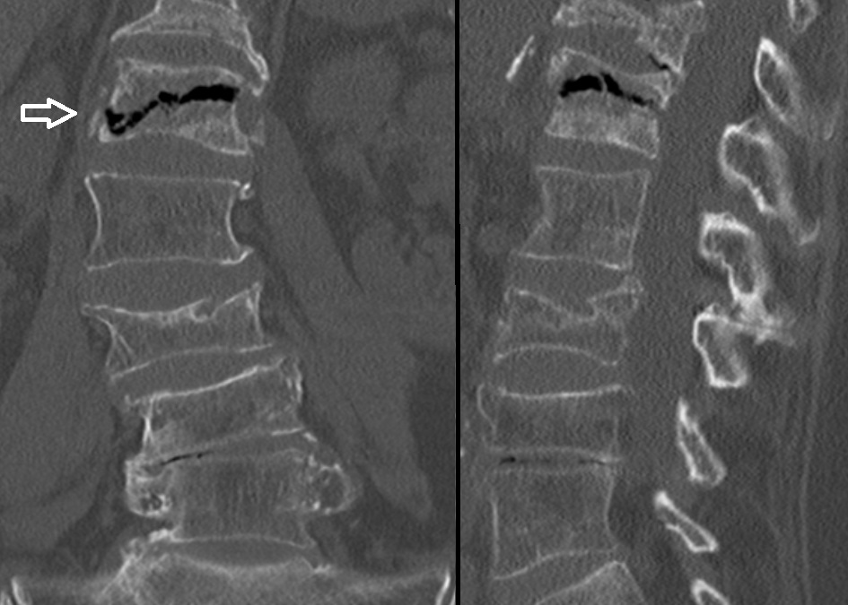
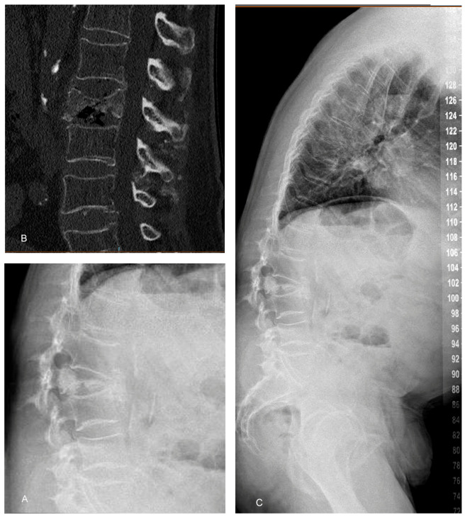
Gas-filled intravertebral vacuum cleft on coronal and sagittal CT. 18 / 60~1 min · at 17:35Mechanism & classificationMost osteoporotic fractures heal with residual deformity and the pain subsides over weeks. The important minority do not.
Tsujio's prospective multicentre study followed 350 conservatively treated patients and found a nonunion rate of 13.5% at six months, defined by a persistent intravertebral cleft. The predictors on the initial films were a thoracolumbar level, a middle-column injury, and specific T2 patterns: a confined area of high signal, or diffuse low signal Tsujio 2011.
The intravertebral cleft, the vacuum sign, is the radiographic marker of a fracture that is still moving. McKiernan found it in every mobile fracture McKiernan 2003, and Ito found it in 79% of delayed collapses before the neurological deficit developed Ito 2002.
Kümmell disease is the eponym for delayed post-traumatic collapse, attributed to avascular necrosis of the vertebral body, presenting weeks to months after a minor fracture with worsening pain, progressive kyphosis, and sometimes a deficit Freedman 2009.
Muratore's systematic review pulled together the predictors of conservative failure: the cleft and the hypointense MRI patterns predict collapse; a middle-column injury predicts nonunion; and retropulsion occupying more than 42% of the canal or more than 15 degrees of change in the wedge angle between positions predicts neurological impairment Muratore 2018. These are the patients who need a surgeon's eye and closer follow-up from the outset.
Pearl: A cleft on the radiograph or CT is a fracture that has not healed. Treat it as a progression risk, not as a curiosity.Refs: Tsujio et al., Spine (Phila Pa 1976), 2011 · McKiernan et al., J Bone Miner Res, 2003 · Ito et al., Spine J, 2002 · Freedman & Heller, J Am Board Fam Med, 2009 · Muratore et al., Eur Spine J, 2018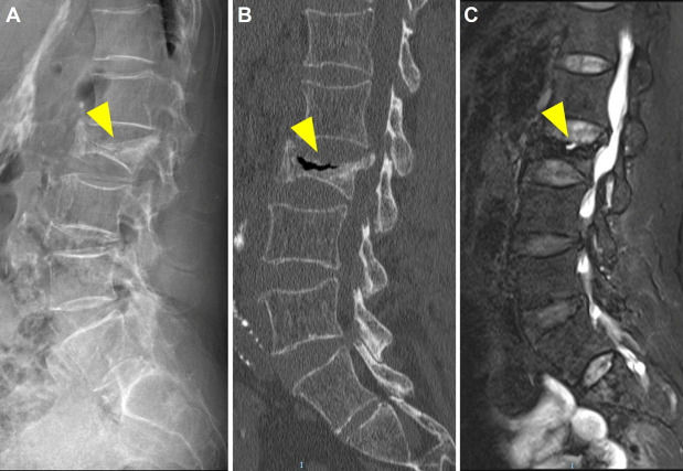
Kümmell disease
19 / 60~1 min · at 18:15Mechanism & classificationThe full picture of Kümmell disease in one patient: a "minor" fracture months earlier, progressive collapse and kyphosis on the radiograph, a gas-filled cleft on CT, and on MRI fluid within the cleft with the posterior wall beginning to bulge toward the canal. This is the vertebra that will not heal by itself and is on its way to a neurological problem Ito 2002 Freedman 2009.
Image from an open-access case report, reproduced under a Creative Commons non-commercial licence with attribution on the slide.
- 20 / 60~0 min · at 18:25Recognition & imaging
Section three, about nine minutes: the clinical picture, the imaging pathway, and the differential that matters most, malignancy.
What the patient tells you
- Acute, localised midline thoracolumbar pain after minimal or no trauma; worse standing and turning in bed, eased lying flat
- Often no neurological symptoms; radicular pain occasionally; ask specifically about leg weakness, numbness and bladder function
- Loss of height, progressive kyphosis, early satiety and shortness of breath in chronic or multiple fractures
- Percussion tenderness over the spinous process at the fractured level; a painful gap or step suggests a more severe injury
- Risk profile: age, prior fragility fracture, glucocorticoids, low weight, smoking, alcohol, and the medications and diseases that weaken bone
- Red flags for another diagnosis: fever, weight loss, night pain, known malignancy, age under 50, pain above T5, or a deficit
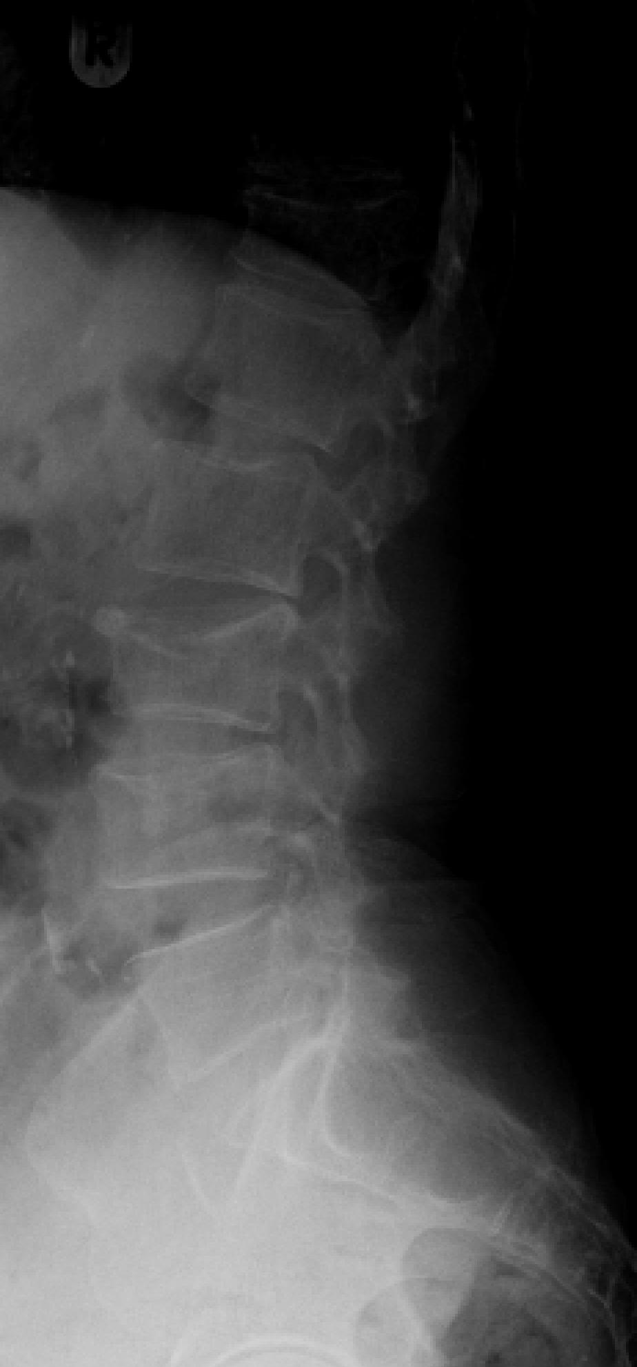
Lateral lumbar radiograph: L3 compression fracture. 21 / 60~1 min · at 19:40Recognition & imagingThe history is usually simple. Sudden midline back pain in an older person after a trivial load, worst on standing and turning, better lying flat. Many patients localise the pain precisely with one finger. Ask about the events of the preceding weeks; a "back strain" three weeks ago may have been the fracture.
Ask the neurological questions directly, because most patients will not volunteer them: leg weakness, numbness, urinary retention or incontinence. Deficits are rare with simple compression fractures, about 3% of inpatients in the German series Schnake 2022, but they change everything.
In the chronic picture, look for height loss, thoracic kyphosis with the rib cage settling onto the pelvis, early satiety, and breathlessness, which follow from the vital capacity data we saw earlier Schlaich 1998.
On examination, percuss the spinous processes; focal tenderness at the fracture level is typical. A palpable gap or step between spinous processes is a posterior ligamentous injury until proven otherwise.
The risk profile tells you how likely osteoporosis is, and the red flags tell you when the fracture might not be osteoporotic: constitutional symptoms, a known cancer, an unusually young patient, an unusual level above T5, or a neurological deficit all push you toward MRI and a broader workup. The ACR appropriateness criteria list exactly these scenarios Expert 2023.
- 22 / 60~1 min · at 20:55Recognition & imaging
The differential in one slide. The dangerous miss is a malignant collapse labelled as osteoporosis, which happens because both are common in the same age group; the spine is the third commonest site of metastasis and myeloma classically presents as a "compression fracture" Expert 2023.
The MRI features come from two classic series. Jung compared 27 metastatic with 55 acute osteoporotic fractures: metastasis was suggested by a convex posterior border, abnormal signal in the pedicle or posterior elements, an epidural or paraspinal mass and other lesions; osteoporosis by a low-signal band on T1 and T2, spared normal marrow elsewhere in the body, a retropulsed posterior fragment, and multiple fractures Jung 2003. Cuénod's series of 93 acute collapses found the same, and added that preserved T1 signal and band-like enhancement favour osteoporosis while inhomogeneous enhancement favours malignancy Cuénod 1996.
Infection is disc-centred and destroys end plates; osteoporotic fractures spare the disc. High-energy trauma follows trauma rules whatever the bone density. Kümmell and nonunion we have covered. And the ankylosed spine, in ankylosing spondylitis or diffuse idiopathic skeletal hyperostosis, fractures like a long bone: a transverse fracture through the fused column that is unstable and often invisible on the first radiograph; the DGOU recommendations treat it as a separate category Blattert 2018.
Bloods are cheap: calcium, phosphate, alkaline phosphatase, full blood count, renal function, and a protein electrophoresis with free light chains if there is any doubt.
- 23 / 60~1 min · at 22:20Recognition & imaging
The pathway. Start with standing radiographs of the whole thoracolumbar spine, because the deformity is often understated on supine films and because multiple levels are common; the DGOU recommendations specify standing images whenever possible Blattert 2018. Compare with any old films you can find; that is the cheapest way to date a fracture. Mehta's series is the reason for the standing film: from supine to erect the Cobb angle went from 11 to 18 degrees, anterior compression from 34 to 46%, and management changed to surgery in a quarter of patients Mehta 2004.
MRI is the decision-making study. Bone-marrow edema on STIR or fat-saturated T2 tells you the fracture is acute, which matters for augmentation eligibility and for attributing the pain to the fracture. MRI also shows the posterior wall, the marrow pattern that separates osteoporosis from malignancy, the ligaments, and any canal compromise. The multispecialty pathway published by Hirsch's panel and the ACR appropriateness criteria both make MRI the preferred advanced study Hirsch 2018 Expert 2023. Get it when the age of the fracture is uncertain, when there are red flags or a deficit, and when a treatment decision depends on it.
CT is for bone detail, and the threshold should be low: Ballock showed that plain radiographs misclassify a quarter of thoracolumbar fractures, almost all bursts read as compression fractures Ballock 1992. The radiographic clues are widening of the interpedicular distance on the AP film and a change in the posterior vertebral body angle, but if the posterior wall is in doubt, get the CT. CT also defines a cleft, an ankylosed spine, and substitutes when MRI is impossible. And image the whole spine when you image: Green's whole-spine MRI series found a second injured level in 77% and non-contiguous fractures in about a third Green 2004; in an osteoporotic patient with multiple deformities, MRI is also how you pick the painful level. And any CT that includes the lumbar spine gives you a free bone density estimate: Schreiber showed that vertebral Hounsfield units correlate with DXA and with compressive strength Schreiber 2011; the ISCD endorses opportunistic CT as a screening tool Anderson 2019.
For the typical fracture with no red flags, treat and repeat standing films at two to four weeks, then six and twelve weeks, looking for progression, and upgrade the OF type if it progresses Blattert 2018.
If the radiograph is normal and the pain persists, remember OF1: the painful vertebra with edema and no deformity, which only MRI shows.
And every branch ends in the same place: the bone-health work-up.
Refs: Mehta et al., Spine (Phila Pa 1976), 2004 · Blattert et al., Global Spine J, 2018 · Hirsch et al., Spine J, 2018 · Green & Saifuddin, Skeletal Radiol, 2004 · Ballock et al., J Bone Joint Surg Br, 1992 · Schreiber et al., J Bone Joint Surg Am, 2011 · Expert et al., J Am Coll Radiol, 2023 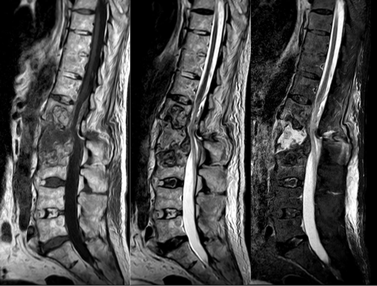
STIR dates the fracture
24 / 60~1 min · at 23:00Recognition & imagingThe same fracture on three sequences. On T1 the fractured body is dark, because edema replaces the bright fatty marrow; on T2 it is subtle; on STIR, which suppresses fat, the edema lights up. That STIR signal is what tells you the fracture is acute and is the pain generator, and its absence in a deformed vertebra tells you the fracture is old. This particular fracture also shows disruption through the posterior elements, a reminder that MRI shows the ligaments too.
Image by Hellerhoff, Wikimedia Commons, Creative Commons Attribution-ShareAlike.
- 25 / 60~1 min · at 24:00Recognition & imaging
A reference table you can photograph. The features come from Jung's Radiographics review and Cuénod's Radiology series Jung 2003 Cuénod 1996.
The two most reliable pointers to malignancy are a convex, bulging posterior cortex and involvement of the pedicles or posterior elements; the two most reliable pointers to osteoporosis are a retropulsed posterior fragment with an otherwise straight cortex, and a band-like low-signal pattern with preserved normal marrow elsewhere. Soft-tissue mass means tumour until proven otherwise. A fluid sign or vacuum cleft is essentially never seen in tumour.
Jung's warning is worth repeating: the signal on a plain fast spin-echo T2 without fat suppression is not helpful. Ask for STIR.
If there is genuine doubt and the patient is going to have an augmentation, send a core biopsy from the same trocar. If they are not, biopsy before you treat.
Refs: Jung et al., Radiographics, 2003 · Cuénod et al., Radiology, 1996 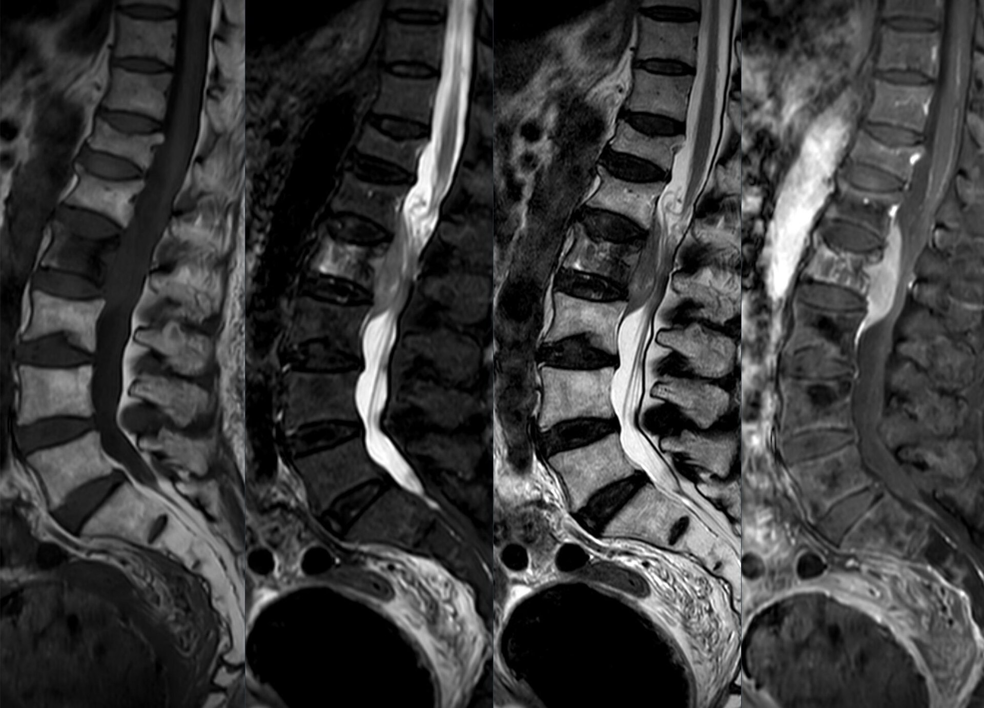
The collapse that is not osteoporosis
26 / 60~1 min · at 24:40Recognition & imagingPut the table into practice. Same age group, same "L2 compression fracture" on the referral. On T1 the whole vertebral body is dark with none of the normal marrow spared; the posterior border is convex and pushes into the canal; and after contrast the body enhances avidly and inhomogeneously. Every column of the malignant side of the table is here Jung 2003 Cuénod 1996. This patient needs staging and a biopsy, not a kyphoplasty.
Image by Hellerhoff, Wikimedia Commons, Creative Commons Attribution-ShareAlike.
- 27 / 60~1 min · at 25:40Recognition & imaging
Twenty seconds for the audience.
The answer is A. She has a dated, typical wedge fracture, neurologically normal, without red flags; that is a fracture to treat and follow with standing radiographs. MRI adds nothing to the immediate decision unless augmentation is being considered or the picture changes. A bone scan is an alternative only when MRI is impossible and the age is uncertain. Flexion-extension films of an acute fracture are painful and unhelpful.
Note the caveat: if she fails to improve, or the follow-up films show progression, the OF type is upgraded and the MRI becomes appropriate.
Refs: Hirsch et al., Spine J, 2018 · Expert et al., J Am Coll Radiol, 2023 · Blattert et al., Global Spine J, 2018 - 28 / 60~0 min · at 25:50Nonoperative care
Section four, about six minutes. This is the treatment most patients get, and the evidence base is thinner than most people assume.
- 29 / 60~1 min · at 27:10Nonoperative care
Nonoperative care is a package, not an absence of treatment.
Analgesia: scheduled paracetamol, a short course of a non-steroidal if renal function and gastric risk allow, and short-acting opioids used sparingly and briefly in this population. Calcitonin is the one drug with trial evidence for the acute pain of vertebral fractures: Knopp-Sihota's meta-analysis of 13 trials found a mean reduction of 3.4 points at rest within a week, and no benefit for chronic fracture pain Knopp-Sihota 2012. The 2010 AAOS guideline gave a moderate recommendation for four weeks of calcitonin Esses 2011. Availability and cost vary; nasal calcitonin is off the market in some regions.
Mobilisation is the treatment. A day or two of rest for pain control is reasonable; prolonged bed rest costs bone and muscle and raises thromboembolic and pulmonary risk in exactly the patients who can least afford it. Physiotherapy teaches safe movement, hip-hinging instead of spinal flexion, and builds back-extensor strength. The 2023 international consensus and the ASBMR task force both endorse exercise for pain, mobility and fear of falling, while judging the evidence that it prevents fractures insufficient Ponzano 2023 Ebeling 2019. The one positive trial is worth knowing: Sinaki's two-year back-extensor programme in 50 postmenopausal women, followed for ten years, was associated with vertebral fractures in 1.6% of vertebrae against 4.3% in controls, a 2.7-fold difference, with stronger extensors and higher bone density Sinaki 2002. Small, but it is the basis of the extension-biased programme physiotherapists teach.
Two things get forgotten. Nutrition: thoracic kyphosis compresses the abdomen, early satiety and weight loss are common, and protein and vitamin D intake matter for healing. And follow-up: standing radiographs at two to four, six and twelve weeks, with escalation if pain fails to settle or the deformity progresses Blattert 2018.
Set expectations. In the randomised trials the conservative arms improved substantially: in Rousing's trial pain fell from about 8 to 2 in both arms by three months Rousing 2009, and in Vertos II 53% of screened patients had spontaneous pain relief before they could even be randomised Klazen 2010. Most patients get better; our job is to identify the ones who will not.
Refs: Knopp-Sihota et al., Osteoporos Int, 2012 · Ponzano et al., Osteoporos Int, 2023 · Sinaki et al., Bone, 2002 · Blattert et al., Global Spine J, 2018 · Rousing et al., Spine (Phila Pa 1976), 2009 · Klazen et al., Lancet, 2010 - 30 / 60~2 min · at 29:00Nonoperative care
Bracing is the intervention everyone prescribes and almost nobody has tested well.
Kim's randomised non-inferiority trial assigned 60 patients with a single-level fracture within three days to no brace, a soft brace, or a rigid brace. At twelve weeks the Oswestry scores were 36, 38 and 34, well within the ten-point non-inferiority margin; pain and the progression of anterior compression were the same in all three groups Kim 2014. Kato's multicentre Japanese trial randomised 284 women to a rigid or a soft brace for twelve weeks and found no difference in vertebral compression at 48 weeks, in pain, or in quality of life Kato 2019.
Pfeifer's trial is the one usually cited in favour of bracing, and it is a different question: a thoracolumbar orthosis worn for six months by women with established kyphosis improved extensor strength, reduced kyphosis and reduced pain Pfeifer 2004. That is an exercise-and-posture device in chronic patients, not acute fracture immobilisation.
Rzewuska's systematic review found low-quality evidence that orthoses beat nothing for medium-term pain and disability Rzewuska 2015; Hofler's review concluded there is no compelling evidence that a rigid brace is superior to a soft brace or no brace Hofler 2020; and the 2023 DGOU review concluded that no superiority for any orthosis could be shown and that, where compared, non-rigid orthoses were favoured Pieroh 2023. The ASBMR task force found it unclear whether bracing improves function, disability or quality of life Ebeling 2019.
So: what the evidence shows is that a rigid brace does not prevent collapse or improve outcomes compared with a soft brace or none. How this affects decisions: stop prescribing a rigid TLSO by reflex, particularly in a frail patient who will not tolerate it and may not mobilise in it. If the patient wants support, a soft or semi-rigid orthosis is reasonable for comfort and confidence. Reserve rigid bracing for a specific surgical plan, for instance an OF3 fracture the surgeon is watching.
Pearl: In two randomised trials, no brace was as good as a rigid brace. The brace is for comfort and confidence, not for preventing collapse. - 31 / 60~1 min · at 30:00Nonoperative care
Two lists: what on the first images should make you worry, and what during follow-up should make you act.
The initial imaging predictors come from Tsujio's prospective study and Muratore's review: a thoracolumbar level, middle-column involvement, the specific T2 and T1 patterns, a cleft or fluid sign, and substantial retropulsion Tsujio 2011 Muratore 2018. A patient with two or more of these deserves earlier follow-up and, in most centres, a surgical opinion from the outset.
The follow-up signs are clinical and radiographic: pain that is not settling by four to six weeks, progressive collapse on standing films with an upgraded OF type, dynamic instability, any new neurological finding, and the patient who simply cannot mobilise. The German inpatient data are sobering: 37% of admitted patients could not be mobilised despite WHO step II analgesia Schnake 2022. That patient is at risk of every complication of immobility and is a candidate for augmentation.
Ito's series is the reminder of what late failure looks like: delayed collapse with a neurological deficit, with the cleft visible in most cases before the deficit appeared Ito 2002.
Refs: Tsujio et al., Spine (Phila Pa 1976), 2011 · Muratore et al., Eur Spine J, 2018 · Ito et al., Spine J, 2002 · Blattert et al., Global Spine J, 2018 - 32 / 60~0 min · at 30:10Vertebral augmentation
Section five, the controversy: about eleven minutes, most of it on the evidence slide.
- 33 / 60~1 min · at 31:25Vertebral augmentation
The two procedures, side by side.
Both start the same way: a cannula through the pedicle, or just lateral to it, into the vertebral body under fluoroscopic guidance, with the patient prone and sedated or anaesthetised. Vertebroplasty injects polymethylmethacrylate cement in a low-viscosity state directly into the fractured trabecular bone; the fill is under pressure, which is why leakage is more common. Kyphoplasty adds a step: a balloon is inflated in the body to create a cavity and to push the end plates apart, restoring some height if the fracture is still mobile; the balloon is withdrawn and thicker cement fills the cavity at lower pressure.
The mechanism of pain relief is thought to be stabilisation of the fracture micromotion, with a possible contribution from thermal and chemical effects on nociceptors; the sham-controlled trials that we will discuss raise the question of how much is placebo and how much is the periosteal anaesthetic.
Newer implantable devices, titanium jacks and vertebral body stents, aim to hold height after the balloon is removed. In the SAKOS randomised trial of 141 patients, the titanium device was non-inferior to balloon kyphoplasty on the composite endpoint, restored more midline height, and had fewer adjacent fractures, 13% versus 27% Noriega 2019. The ASBMR task force found insufficient evidence to prefer stenting over kyphoplasty Ebeling 2019.
Refs: Noriega et al., Spine J, 2019 - 34 / 60~3 min · at 34:25Vertebral augmentation
This is the evidence slide, and it is worth being precise because the trials genuinely disagree.
Two sham-controlled trials in the New England Journal in 2009 changed the field. Buchbinder randomised 78 patients with fractures of less than twelve months to vertebroplasty or a sham procedure and found no advantage at any time point; both groups improved, with three-month pain reductions of 2.6 and 1.9 Buchbinder 2009. INVEST randomised 131 patients and found no difference in disability or pain at one month, though 51% of controls crossed over versus 13% of the vertebroplasty group Kallmes 2009. In both, the sham included infiltration of the periosteum with local anaesthetic.
Vertos II, open-label, took a more selected population: fractures of six weeks or less, edema on MRI, pain of five or more. Of 431 eligible patients, 53% had spontaneous relief during assessment and were never randomised; among the 202 who still had pain, vertebroplasty gave 2.6 points more relief at one month and 2.0 at one year Klazen 2010. That screening finding is important in itself: half of these patients get better on their own within weeks.
VAPOUR tried to answer the critics with a sham-controlled trial of very acute, very painful fractures: under six weeks, pain of seven or more. The primary outcome, pain below four at fourteen days, was achieved by 44% with vertebroplasty versus 21% with placebo Clark 2016. The sham did not include periosteal anaesthetic, and the trial was supported by an industry grant.
VERTOS IV then repeated the sham design in 180 acute patients, with periosteal anaesthetic in both arms, and found differences of 0.2 to 0.45 points on a ten-point scale across twelve months, none significant Firanescu 2018.
For kyphoplasty, FREE randomised 300 patients to kyphoplasty or non-surgical care and found a 5.2-point advantage in the SF-36 physical component at one month; by twelve months the quality-of-life difference was no longer significant, though back pain remained modestly better at two years, and new fractures were similar, 47.5% versus 44.1% Wardlaw 2009 Boonen 2011.
Most recently VERTOS V addressed chronic fractures, more than three months old but with persisting edema, against an active control of anaesthetic infiltration: at twelve months pain was 3.9 versus 5.1, quality of life was better, disability was not different Carli 2023.
The pooled analyses reflect the split. Láinez's meta-analysis of 14 trials found vertebroplasty clearly better than conservative care, slightly better than a skin-only placebo, and no better than a sham that infiltrated the periosteum, with the benefits largest in fractures under six weeks Láinez 2021. Mattie found a pooled effect close to the 1.5-point minimal clinically important difference Mattie 2016; Halvachizadeh found 1.3 points short-term and no increase in adjacent fractures Halvachizadeh 2021. The Cochrane review, using the sham trials as the primary comparison, concluded that vertebroplasty provides no clinically important benefit over placebo, with a 0.7-point pain difference at one month, and that harms cannot be excluded Buchbinder 2018.
Read the two summary boxes. What the evidence shows: against a sham that anaesthetises the periosteum, no important benefit; against conservative care, faster pain relief with the largest effects in severe, recent fractures; effects fade by a year; no convincing effect on the bone or on subsequent fractures. How this affects decisions: augmentation is not routine care. It is a reasonable option for the patient with an MRI-confirmed acute fracture and severe pain who has failed adequate analgesia and cannot function, particularly the hospitalised patient who cannot mobilise, after a frank discussion. It is a pain procedure; the bone still needs treating.
- 35 / 60~1 min · at 35:25Vertebral augmentation
The guidelines disagree, and the disagreement tracks the specialty of the authors.
The 2010 AAOS guideline made a strong recommendation against vertebroplasty on the basis of the two sham trials, and a weak recommendation that kyphoplasty is an option for neurologically intact patients Esses 2011. The Cochrane review and the ASBMR task force, dominated by rheumatologists and bone physicians, concluded that vertebroplasty has no clinically important benefit over sham, that the evidence for kyphoplasty is insufficient, and that routine use is not supported, while stressing that patients must be fully informed Buchbinder 2018 Ebeling 2019.
The multispecialty pathway from Hirsch's panel, largely interventionalists and surgeons using the RAND appropriateness method, found augmentation appropriate in patients with positive advanced imaging and worsening symptoms, or with two to four unfavourable conditions such as progressive height loss or severe functional impact Hirsch 2018. The ACR criteria rate scenarios and place MRI before treatment, with augmentation appropriate for refractory painful acute fractures Expert 2023. The 2025 NASS guideline addresses 29 questions; the summary is in The Spine Journal and the graded recommendations are on the NASS website Cho 2025.
My reading for a mixed audience: the sham trials set a ceiling on the benefit for the average patient; the appropriateness pathways describe the subgroup in whom it is reasonable to offer it. Neither justifies augmentation as a reflex, and none of them treats the underlying bone.
Refs: Esses et al., J Am Acad Orthop Surg, 2011 · Buchbinder et al., Cochrane Database Syst Rev, 2018 · Ebeling et al., J Bone Miner Res, 2019 · Hirsch et al., Spine J, 2018 · Expert et al., J Am Coll Radiol, 2023 · Cho et al., Spine J, 2025 - 36 / 60~1 min · at 36:25Vertebral augmentation
You will hear the mortality argument, so here it is with its counterweight.
Edidin analysed 858,978 Medicare vertebral fracture patients: those who had augmentation had a four-year survival of 61% versus 50% for nonoperated patients, a 37% lower adjusted hazard, and kyphoplasty patients did better than vertebroplasty patients Edidin 2011. Chen found the same pattern in 68,752 patients: three-year survival 42% nonoperative, 50% vertebroplasty, 60% kyphoplasty, with fewer pneumonias and pressure ulcers in the kyphoplasty group, and the authors themselves flagged selection bias Chen 2013. Hinde's meta-analysis of more than two million patients pooled a hazard ratio of 0.78 Hinde 2020.
The counterweight is the 2025 meta-analysis by Wiedl, which included the two randomised trials and eleven studies with hazard ratios: the pooled hazard ratio was 0.91 with a confidence interval crossing one, and the randomised data showed no mortality difference Wiedl 2025. The obvious confounder is that the patients selected for a procedure are the ones fit enough to have one.
The honest conclusion: the registry survival advantage is real as an association and unproven as a causal effect. It should not be used to sell the procedure, but it is a fair reminder that immobility kills these patients and that the goal of any treatment is to get them upright.
- 37 / 60~1 min · at 37:40Vertebral augmentation
Selection is where the trials become practice.
A reasonable candidate has an MRI-confirmed acute or unhealed fracture with edema at the level that hurts; severe pain despite a proper trial of analgesia, or an inability to mobilise, particularly the inpatient who cannot get out of bed; and a fracture that is recent, because the largest effects in the trials were in fractures under six weeks, although the appropriateness panel considered edema more relevant than the calendar Láinez 2021 Hirsch 2018. Exclusions are the obvious ones: infection, uncorrectable coagulopathy, a malignant fracture without a biopsy plan, and a burst with retropulsion and a deficit, which is a surgical problem.
Complications. Hulme's review of 69 studies put cement leakage at 41% of vertebrae after vertebroplasty and 9% after kyphoplasty Hulme 2006; Taylor's kyphoplasty meta-analysis found 8% leakage with 0.09% symptomatic, 11% new fractures, and rare cord compression, pulmonary embolism and death Taylor 2007. Most leakage goes into the disc, the paravertebral veins or the epidural space and is silent; symptomatic leakage with cord compression, pulmonary cement embolism and osteomyelitis are all reported in the trials and reviews Buchbinder 2018. Trout's series found new fractures in 20% of vertebroplasty patients, 41% of them at adjacent levels and occurring sooner, at a median of 55 days Trout 2006; Fribourg found new fractures in 26% within eight months of kyphoplasty, and every fracture in the first two months was adjacent Fribourg 2004; Lin showed that cement leaking into the disc raised the adjacent fracture rate from 12% to 58% Lin 2004, so keep the cement out of the disc, keep the volume modest, and do not augment adjacent levels prophylactically, for which the chapters found no evidence of benefit Schmidt 2011; the randomised data, however, do not show more new fractures than with conservative care Halvachizadeh 2021, and FREE found similar rates in both arms Boonen 2011. In the Medicare data kyphoplasty patients were slightly more likely to have another augmentation Chen 2013. And do not forget that this is a prone procedure under sedation in an eighty-year-old with a kyphotic chest. The risk of an epidural leak rises with a breached posterior wall, low-viscosity cement under pressure and operator inexperience, which is one more reason the posterior wall matters in selection; cord compression from an epidural leak is a surgical emergency Buchbinder 2018. The osteoporotic-surgery chapter puts it plainly: these are not minor interventions to be done without an anaesthetist and a spine surgeon on call Schmidt 2011.
On selection by fracture age, the chapters are more permissive than the trials: Kasperk's prospective controlled cohort treated fractures older than twelve months and still found better height, pain and function than conservative care at six months Kasperk 2005, and the compression-fracture chapter concludes that fracture age is not a critical factor and edema is not a prerequisite McCall 2011. VERTOS V, on the evidence slide, says the same for chronic fractures with edema. Persistent edema and pain at the level are the criteria, and the six-week figure is a guide, not a gate. Note also how the evidence moved: the 2011 chapters, written on the open-label trials, call the evidence for augmentation strong Schmidt 2011; the sham trials of 2009 and the ones that followed are why this lecture calls it an option for selected patients.
Refs: Láinez et al., Eur Radiol, 2021 · Hirsch et al., Spine J, 2018 · Kasperk et al., J Bone Miner Res, 2005 · Hulme et al., Spine (Phila Pa 1976), 2006 · Taylor et al., Eur Spine J, 2007 · Buchbinder et al., Cochrane Database Syst Rev, 2018 · Fribourg et al., Spine (Phila Pa 1976), 2004 · Lin et al., AJNR Am J Neuroradiol, 2004 · Halvachizadeh et al., JBJS Rev, 2021 · Chen et al., J Bone Joint Surg Am, 2013 · Schmidt et al., In: Vaccaro AR, Fehlings MG, Dvorak MF, eds. Spine and Spinal Cord Trauma: Evidence-Based Management. Thieme, 2011 - 38 / 60~1 min · at 38:40Vertebral augmentation
Twenty seconds.
B is the answer, and the wording matters: reasonable, with an honest discussion. This is the patient the pathway calls appropriate and the patient VAPOUR enrolled Hirsch 2018 Clark 2016. The sham trials tell us the average effect is small, not that the procedure is contraindicated Buchbinder 2018. A rigid brace will not fix his problem. And the mortality figure is an association from registry data that the meta-analysis including randomised trials did not confirm Wiedl 2025.
Refs: Hirsch et al., Spine J, 2018 · Clark et al., Lancet, 2016 · Buchbinder et al., Cochrane Database Syst Rev, 2018 · Wiedl et al., Dtsch Arztebl Int, 2025 - 39 / 60~0 min · at 38:50Burst fractures & surgery
Section six, about nine minutes. This is the part of the talk for the orthopaedic residents, but everyone needs the first slide: the fractures that must reach a surgeon.
The osteoporotic fracture that is not benign
- Burst fracture: posterior wall involved (OF3, AOSpine A3–A4) or vertebral frame lost (OF4) Schnake 2018 Vaccaro 2013. Retropulsion alone in an intact patient is not an indication: it remodels Wood 2003 Cantor 1993
- Neurological deficit: rare (about 3% of inpatients) but an indication for decompression and stabilisation Schnake 2022 Blattert 2018
- Delayed collapse with a cleft (Kümmell) and progressive kyphosis; retropulsion over 42% of the canal or over 15° dynamic change predicts deficit Muratore 2018
- Distraction or rotation injuries (OF5, AOSpine B or C, TLICS ≥ 5), and any fracture in an ankylosed spine: 67% of AS fractures present with a deficit and 3-month mortality is about 18% Westerveld 2009 Blattert 2018
- Multiple adjacent fractures with sagittal decompensation: the patient who can no longer stand upright
- Painful nonunion that has failed conservative care and is not suitable for augmentation alone
L2 osteoporotic fracture: segmental kyphosis, vacuum cleft and posterior wall involvement on CT. 40 / 60~1 min · at 40:00Burst fractures & surgeryEveryone in the room should know this list, because these are the patients who must reach a spine surgeon and must not be discharged with analgesics.
Burst fractures: once the posterior wall is involved, OF3, and certainly once the vertebral frame has failed, OF4, the fracture can retropulse into the canal and can collapse further under load Schnake 2018. Neurological deficits are uncommon, about 3% in the German inpatient series, but they are the clearest indication for decompression and stabilisation Schnake 2022 Blattert 2018.
Delayed collapse with a cleft, the Kümmell picture, with progressive kyphosis, and the imaging thresholds Muratore's review associated with neurological impairment: retropulsion beyond 42% of the canal and more than 15 degrees of dynamic wedge change Muratore 2018.
OF5 injuries with distraction or rotation, and any fracture through an ankylosed spine, are unstable; the DGOU treats ankylosing spondylitis fractures as a separate category needing stabilisation Blattert 2018. Westerveld's systematic review of 345 ankylosing spondylitis and 55 DISH patients with spinal fractures found a neurological deficit on admission in 67% and 40%, complications in half, and 3-month mortality of 18 to 20%, mostly from recumbency Westerveld 2009. The chapters add that these fractures are missed in a fifth, deteriorate before treatment in about one in seven, and belong in a category of their own: three levels above and below, percutaneous when possible, and a low threshold for MRI because epidural haematoma is common Williams 2011.
A word on the canal, because residents are still taught the historic thresholds of 30 degrees of kyphosis, 50% height loss and 50% canal compromise. Those were pre-MRI surrogates for posterior ligament disruption. In a neurologically intact patient with an intact posterior ligamentous complex, canal compromise on its own is not an operative indication: in Wood's randomised trial the retropulsed fragment remodelled from 39% to 22% of the canal with surgery and from 34% to 19% without, and Cantor's early-ambulation series showed the same resorption with no neurological deterioration Wood 2003 Cantor 1993. Muratore's 42% threshold on the previous bullet is a different situation: progressive osteoporotic collapse with a cleft, where the fragment is moving, not a stable traumatic burst. Do not operate on a number; operate on instability, progression and the examination.
Then the two chronic problems: the patient with multiple adjacent fractures who has lost sagittal balance and cannot stand upright, and the painful nonunion that has exhausted conservative care and is not a candidate for augmentation alone.
Pearl: The posterior wall and the neurological examination decide whether this is a pain problem or a surgical problem.Refs: Schnake et al., Global Spine J, 2018 · Vaccaro et al., Spine (Phila Pa 1976), 2013 · Wood et al., J Bone Joint Surg Am, 2003 · Cantor et al., Spine (Phila Pa 1976), 1993 · Schnake et al., Eur J Trauma Emerg Surg, 2022 · Blattert et al., Global Spine J, 2018 · Muratore et al., Eur Spine J, 2018 · Westerveld et al., Eur Spine J, 2009- 41 / 60~1 min · at 41:10Burst fractures & surgery
Before the osteoporosis-specific operations, the baseline every resident should know: the trauma literature on the burst fracture without a deficit, reviewed in the Vaccaro chapter Sekhon 2011.
Wood's randomised trial is the anchor: 47 stable burst fractures without deficit, followed for a mean of 44 months. Kyphosis behaved the same in both arms, the canal remodelled the same in both arms, from about 35 to 40% down to about 20%, pain and return to work were similar, and the nonoperative patients reported less disability and had fewer complications Wood 2003. Shen's prospective comparison found that surgery buys less pain for three months and better function for six, then nothing, at four times the cost, while the retropulsed fragment in the braced patients resorbed from 34 to 15% of the canal Shen 2001. Siebenga's multicentre trial is the dissent, with better kyphosis, pain and return to work after surgery, in 34 patients with mixed fracture types Siebenga 2006. Bailey's equivalence trial then removed the brace: for A3 bursts, function at three months was the same with and without a thoracolumbosacral orthosis Bailey 2014; that extends what Kim and Kato showed for compression fractures to the burst. And Cantor's series is the one to quote when someone points at the fragment in the canal: it resorbs, and nobody deteriorated Cantor 1993.
The Spine Trauma Study Group's conclusion in the chapter is that a brace for six to twelve weeks with limited bed rest is a reasonable option, that anterior and posterior approaches are both acceptable when surgery is chosen, and that in the elderly with poor bone quality the posterior approach is favoured Sekhon 2011. Everything that follows in this section is about the ways osteoporotic bone breaks those rules: it collapses under load, it clefts, and it does not hold screws.
Refs: Wood et al., J Bone Joint Surg Am, 2003 · Shen et al., Spine (Phila Pa 1976), 2001 · Siebenga et al., Spine (Phila Pa 1976), 2006 · Bailey et al., Spine J, 2014 · Cantor et al., Spine (Phila Pa 1976), 1993 · Sekhon, In: Vaccaro AR, Fehlings MG, Dvorak MF, eds. Spine and Spinal Cord Trauma: Evidence-Based Management. Thieme, 2011 - 42 / 60~1 min · at 42:25Burst fractures & surgery
The biomechanical argument behind every osteoporotic fixation principle, drawn from the vertebroplasty and minimally invasive chapters of the trauma text.
McCormack's load-sharing classification scored comminution, fragment apposition and the amount of kyphosis correction, and predicted which short-segment posterior constructs would fail: a score of seven or more needs the anterior column reconstructed McCormack 1994. In osteoporotic bone almost every burst scores high on comminution, and the correction you achieve is exactly what the bone cannot hold.
Tezeren's randomised comparison is the cleanest demonstration: short-segment versus long-segment fixation for burst fractures, 55% implant failure in the short constructs and none in the long, with identical clinical scores Tezeren 2005. That is the origin of the rule that constructs are longer in this population.
Oner explained the mechanism of recurrent kyphosis with late MRI: ligamentotaxis through the annulus reduces the periphery of the body but leaves the central end plate depressed, the disc creeps into the crater, and the segment settles into kyphosis; it is not degeneration and it is not instability, it is an unsupported anterior column Oner 1998.
So fill the void. Mermelstein's cadaveric study showed transpedicular calcium phosphate cement under the end plate cut the bending moments on the screws by 59% in flexion and raised the stiffness of the construct by 40% Mermelstein 1998; Verlaan's phase II series combined balloon reduction and cement with short-segment screws and restored anterior height to 91% of intact with no fragment displacement Verlaan 2005. Mahar showed that adding screws at the fractured level itself increases torsional stiffness and holds the reduction Mahar 2007. Uchida's clinical series, on the next slide, is the same idea in osteoporotic patients with deficits.
And the invasiveness argument for this age group. Korovessis treated 18 older patients with lumbar burst and severe compression fractures by balloon kyphoplasty plus short percutaneous instrumentation: kyphosis from 16 to 2 degrees, 45 minutes, 70 millilitres of blood loss, no implant failure or loss of correction at 22 months Korovessis 2008. Wild's five-year comparison found percutaneous stabilisation halved blood loss with identical correction and outcome scores Wild 2007. Against that, Rechtine's series of open trauma instrumentation carried a 10% wound infection rate, 41% in patients with complete cord injuries Rechtine 2001. Minimally invasive posterior fixation with augmentation of the index level is the natural construct for an eighty-year-old.
Pearl: In osteoporotic bone the anterior column will not carry the load, so give it something to stand on and do not ask a short construct to do it alone.Refs: McCormack et al., Spine (Phila Pa 1976), 1994 · Tezeren & Kuru, J Spinal Disord Tech, 2005 · Oner et al., J Bone Joint Surg Br, 1998 · Mermelstein et al., Spine (Phila Pa 1976), 1998 · Verlaan et al., Spine (Phila Pa 1976), 2005 · Mahar et al., Spine (Phila Pa 1976), 2007 · Korovessis et al., Spine (Phila Pa 1976), 2008 · Wild et al., Arch Orthop Trauma Surg, 2007 · Rechtine et al., J Orthop Trauma, 2001 - 43 / 60~1 min · at 43:50Burst fractures & surgery
Three families of operation, all supported by retrospective series rather than trials.
Kyphoplasty alone for a burst fracture sounds heretical but has a literature. Chen's systematic review pooled 289 patients with osteoporotic thoracolumbar burst fractures treated by kyphoplasty alone: pain fell from 6.8 to 1.1 and kyphosis from 22 to 3 degrees, at the cost of cement leakage in 8 to 45% and adjacent fracture or relapse in a wide range Chen 2021. The prerequisites are a posterior wall that is displaced but not disrupted, no neurological deficit, and meticulous technique. Many surgeons would call this an OF3 or selected OF4 indication.
Posterior instrumentation is the workhorse for instability, deficit and collapse, and the recurring problem is that the anterior column does not support the construct. Uchida compared short-segment posterior fixation with vertebroplasty of the fractured body, posterior fixation alone, and anterior surgery in 83 patients with collapse and deficit: neurological outcomes were the same, but loss of correction was 4.6 degrees with augmentation versus 8.6 without, equivalent to anterior reconstruction Uchida 2010. The DGOU principles are explicit: stability over motion preservation, cement-augmented or high-purchase screws, and restoration of the individual pre-fracture sagittal profile Blattert 2018. For Kümmell disease, Yu's meta-analysis found that long-segment fixation held the kyphosis better at final follow-up while short-segment fixation saved operative time and blood loss, with similar complications Yu 2022.
Anterior reconstruction addresses the biomechanics directly: the anterior column at the thoracolumbar junction carries most of the load. Kanayama's series of Kaneda reconstructions showed neurological recovery in all patients, but kyphosis drifted back and 19% needed posterior reinforcement Kanayama 2010. Sudo's comparison found similar neurological recovery with anterior and posterior approaches, but 34% respiratory complications with anterior surgery and a need for early posterior reinforcement when bone density was below 0.60 grams per square centimetre or the construct spanned multiple collapses Sudo 2013. For fixed kyphosis with a deficit, Suk showed that a posterior closing-wedge osteotomy matched combined anterior-posterior surgery for neurological improvement with less operative time, blood loss and morbidity Suk 2003.
Two principles for the patient with a deficit, from the chapters. Timing: a progressive deficit is operated on as soon as the patient is resuscitated, but the eighty-year-old on anticoagulants is optimised first; Kerwin's series of early versus late spine fixation found shorter hospital stays with fixation within three days but a mortality trend of 6.9% against 2.5% in the early group Kerwin 2005. Decompression: laminectomy alone is contraindicated, because it does not reach an anterior fragment and it destabilises the segment; decompress from the direction of the compression, or indirectly by reduction, and stabilise Sekhon 2011.
The footnote is the honest summary: no randomised trial exists, and the PubMed search for this lecture found none.
For context, the prospective German inpatient study of 707 patients across 16 trauma centres shows what specialist centres do: mean age 75, mostly A1 and A3 fractures, deficits in 3%, 37% unable to mobilise on step II analgesia, and 82% treated surgically, mostly kyphoplasty alone with hybrid kyphoplasty-plus-screw constructs in about a fifth, invasiveness rising with instability Schnake 2022. Most vertebral fractures never reach an inpatient bed; these are the ones that do.
Refs: Chen et al., Pain Physician, 2021 · Uchida et al., J Neurosurg Spine, 2010 · Blattert et al., Global Spine J, 2018 · Yu et al., Geriatr Orthop Surg Rehabil, 2022 · Kanayama et al., J Spinal Disord Tech, 2010 · Sudo et al., Spine J, 2013 · Suk et al., Spine (Phila Pa 1976), 2003 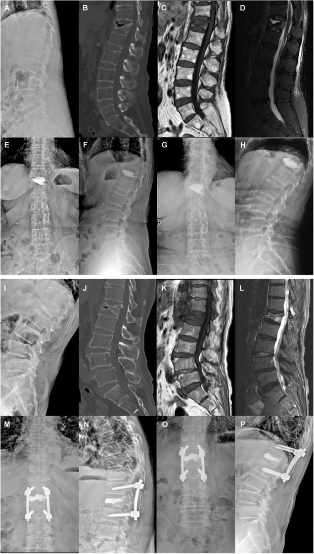
Two answers to the same problem
44 / 60~1 min · at 44:30Burst fractures & surgeryTwo patients with stage 3 Kümmell disease, meaning collapse with posterior wall involvement, from a comparative series. The upper patient was treated with kyphoplasty alone; the lower with short-segment posterior fixation plus vertebroplasty of the collapsed body. The choice turns on the posterior wall, the degree of instability on standing films, the presence of a deficit, and the patient's fitness for an open operation. Uchida's data support augmenting the index level whenever posterior fixation is used, to prevent loss of correction Uchida 2010.
Image from an open-access article, Creative Commons Attribution.
Fixation in osteoporotic bone
- Expect screw pull-out and loosening; plan for it rather than discover it
- Cement-augmented pedicle screws: pooled loosening 2.0%, breakage 0.6%; any cement leakage 21.8% but symptomatic leakage 1.2%, symptomatic pulmonary embolism 0.8% Zhang 2021
- PMMA and calcium-based cements both increase pull-out strength; fenestrated screws and pre-fill techniques perform similarly Elder 2015
- Longer constructs, more fixation points, and hooks or bands to share load; avoid ending a construct at the apex of a kyphosis or next to a fractured level
- Reconstruct the anterior column when it cannot bear load: augmentation of the index vertebra, cage, or strut; PMMA, not calcium phosphate, for stand-alone fill in osteoporotic bone Uchida 2010 Sudo 2013 Schmidt 2011
- Restore the patient's own pre-fracture sagittal profile, not a textbook one; minimise invasiveness in this population Blattert 2018
- Treat the bone: bone-health optimisation improves outcomes and anabolic therapy is favoured around instrumentation Anderson 2019
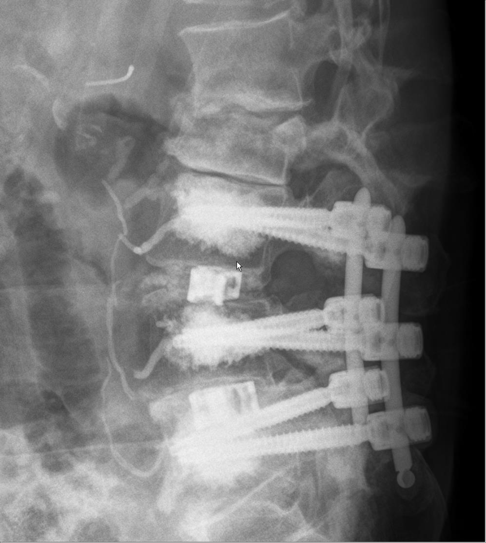
Three-level posterior fixation with cement-augmented pedicle screws. 45 / 60~1 min · at 45:40Burst fractures & surgerySeven principles for the residents.
First, expect fixation failure and design against it. The pedicle screw in osteoporotic bone has a fraction of the pull-out strength of the same screw in young bone.
Cement augmentation of screws is the most-used answer. Zhang's meta-analysis of twenty studies found low rates of screw-related complications, 2% loosening and 0.6% breakage, but cement leakage in a fifth of patients, almost all of it silent; symptomatic leakage was 1.2% and symptomatic pulmonary embolism 0.8% Zhang 2021. Elder's biomechanical review found that PMMA and calcium ceramics both improve fixation and that fenestrated screws and pre-fill techniques are comparable Elder 2015.
Constructs are longer than in young patients, with more fixation points, and hooks or sublaminar bands at the ends to share load. Do not stop a construct at the apex of the kyphosis or adjacent to another fractured level.
The anterior column must bear load or the posterior construct will fail: augment the index vertebra, or place a cage or strut where the body is gone Uchida 2010 Sudo 2013.
The DGOU principles: restore the patient's own pre-fracture sagittal profile, which in a compensated older spine is not a textbook lordosis, and keep the surgery as minimally invasive as the goals allow Blattert 2018.
And treat the bone. The American Orthopaedic Association's bone-health optimisation position recommends assessing every patient over 50, correcting metabolic deficits, and considering delaying elective surgery three months to start treatment; around instrumentation in fragile bone, anabolic agents are the logical choice Anderson 2019.
Refs: Zhang et al., Orthop Traumatol Surg Res, 2021 · Elder et al., Spine J, 2015 · Uchida et al., J Neurosurg Spine, 2010 · Sudo et al., Spine J, 2013 · Schmidt et al., In: Vaccaro AR, Fehlings MG, Dvorak MF, eds. Spine and Spinal Cord Trauma: Evidence-Based Management. Thieme, 2011 · Blattert et al., Global Spine J, 2018 · Anderson et al., J Bone Joint Surg Am, 2019- 46 / 60~0 min · at 45:50Bone health
Section seven, about nine minutes, and the highest-yield section for the patient: secondary fracture prevention, with the drugs by name.
- 47 / 60~1 min · at 46:50Bone health
The treatment gap in four numbers.
Solomon's analysis of nearly 97,000 US hip-fracture patients found that only 28.5% received any osteoporosis medication in the following year, and the rate fell over the decade from 40% to 20% Solomon 2014; the strongest predictor of treatment after the fracture was treatment before it. For vertebral fractures the gap is worse because the fracture itself is not recognised: in Gehlbach's series, 18% of women with a fracture on their chest radiograph received treatment Gehlbach 2000.
Against that, the effect size of treatment is among the largest in medicine: the ASBMR task force summarised the drug trials as a 40 to 70% reduction in subsequent vertebral fractures Ebeling 2019.
And the system fix works. Wu's meta-analysis of 74 controlled studies of fracture liaison services found treatment initiation up by 20 percentage points, adherence up, re-fracture down from 13.4 to 6.4%, and mortality down from 15.8 to 10.4% Wu 2018. If your hospital does not have a fracture liaison service or an Own the Bone programme, this slide is the business case.
- 48 / 60~1 min · at 48:00Bone health
The work-up is short and the orthopaedic surgeon can start it.
First principle from the clinician's guide: a fragility fracture of the spine or hip after age 50 is diagnostic of osteoporosis whatever the T-score says, and it signals imminent risk; a vertebral fracture carries about a five-fold risk of another vertebral fracture and a two- to three-fold risk of other fractures LeBoff 2022. You do not need the DXA to justify treatment, but you need it to stage and to monitor.
DXA of the hip and spine. The 2025 USPSTF statement recommends screening women 65 and over and younger postmenopausal women at increased risk, and finds the evidence insufficient in men US 2025; your patient with a fracture is past screening and into treatment. FRAX gives a ten-year probability of hip and major osteoporotic fracture from clinical risk factors with or without bone density Kanis 2008; the recent fracture increases the true risk beyond the calculator.
The laboratory screen looks for secondary causes: calcium and phosphate, alkaline phosphatase, renal function, vitamin D, thyroid function, a blood count, and a protein electrophoresis with light chains because myeloma hides in this population. Testosterone in men; parathyroid hormone and coeliac serology when the picture suggests it.
If a CT has already been done for any reason, measure the Hounsfield units in the L1 body: Schreiber showed the correlation with DXA and compressive strength, and the ISCD endorses opportunistic CT Schreiber 2011 Anderson 2019.
And the ownership point: the ISCD position says patients with a fracture after 50, inflammatory arthritis, chronic steroids or renal disease should have DXA before orthopaedic surgery Anderson 2019, and the AOA's bone-health optimisation position asks surgeons to assess everyone over 50, correct deficits, and delay elective surgery three months to start treatment when feasible Anderson 2019.
Refs: LeBoff et al., Osteoporos Int, 2022 · US et al., JAMA, 2025 · Kanis et al., Osteoporos Int, 2008 · Schreiber et al., J Bone Joint Surg Am, 2011 · Anderson et al., J Clin Densitom, 2019 · Anderson et al., J Bone Joint Surg Am, 2019 - 49 / 60~1 min · at 49:20Bone health
The antiresorptives, by trade name, with the numbers a patient will ask about.
Alendronate, Fosamax, 70 mg once a week. The Fracture Intervention Trial, in women who already had a vertebral fracture: new morphometric vertebral fractures fell from 15% to 8%, clinical vertebral fractures by more than half, and hip fractures by about half over three years Black 1996. This is the trial that makes bisphosphonates first-line in every guideline, and the generic costs a few dollars a month.
Risedronate, Actonel, 35 mg weekly, or Atelvia, the delayed-release form taken after breakfast. VERT North America: vertebral fractures down 41% over three years, and 65% in the first year, which matters for a patient whose highest-risk period is the next twelve months; nonvertebral fractures down 39% Harris 1999.
Ibandronate, Boniva, monthly tablet or three-monthly injection. BONE showed a 62% reduction in vertebral fractures but no effect on nonvertebral or hip fractures Chesnut 2004; it is a spine-only drug and most guidelines list it behind the other bisphosphonates for that reason.
Zoledronic acid, Reclast, one 5 mg infusion a year. HORIZON: vertebral fractures down 70%, hip fractures down 41%, nonvertebral down 25% Black 2007. For the patient who cannot manage or tolerate a tablet, or who you doubt will take one, this is the bisphosphonate: one infusion, given before discharge if the vitamin D is replete and the eGFR is above 35. Expect an acute-phase reaction after the first dose in about a third; the atrial fibrillation signal was not confirmed in later trials but is on the label.
Denosumab, Prolia, 60 mg under the skin every six months. FREEDOM: vertebral fractures down 68%, hip down 40%, nonvertebral down 20% Cummings 2009. It works in renal impairment, which the bisphosphonates do not. The price is commitment: it is not a drug you can stop, and the slide after next explains why.
Raloxifene, Evista, 60 mg daily. MORE: vertebral fractures down 30%, no effect on hip or nonvertebral fractures, and a three-fold risk of venous thromboembolism Ettinger 1999. It has a place in a younger postmenopausal woman with spine-predominant osteoporosis and a breast cancer concern, and almost no place in the patient in front of you.
Every one of these needs calcium and vitamin D replete first, and the bisphosphonates need a creatinine clearance above about 30 to 35.
Refs: Black et al., Lancet, 1996 · Harris et al., JAMA, 1999 · Chesnut et al., J Bone Miner Res, 2004 · Black et al., N Engl J Med, 2007 · Cummings et al., N Engl J Med, 2009 · Ettinger et al., JAMA, 1999 - 50 / 60~1 min · at 50:40Bone health
The anabolics, which is where the field has moved for the patient with a fresh vertebral fracture.
Teriparatide, Forteo, also sold as Bonsity, recombinant parathyroid hormone 1 to 34, 20 micrograms under the skin every day for up to two years. Forteo is the name you will meet most often in the spine-fusion literature. In the Fracture Prevention Trial, in women with prior vertebral fractures, new vertebral fractures fell from 14% to 5%, a relative risk of 0.35, and nonvertebral fragility fractures were halved Neer 2001. Side effects are nausea, cramps and mild hypercalcaemia. The osteosarcoma boxed warning that limited it to two years lifetime was removed from the US label in 2020 after twenty years of surveillance; the two-year course remains standard. It is contraindicated in Paget disease, open physes, unexplained raised alkaline phosphatase and prior skeletal radiotherapy, and it has no place in a patient with a bone metastasis.
VERO put teriparatide head to head with risedronate in women with severe osteoporosis, at least two moderate or one severe vertebral fracture: new vertebral fractures 5.4% versus 12%, clinical fractures halved Kendler 2018. That is the trial to quote when someone says a bisphosphonate is good enough after a vertebral fracture.
Abaloparatide, Tymlos, a parathyroid hormone-related peptide analogue, 80 micrograms daily. ACTIVE against placebo over 18 months: new vertebral fractures 0.58% versus 4.22%, an 86% reduction, nonvertebral fractures down 43%, with less hypercalcaemia than the teriparatide arm Miller 2016. Efficacy is similar to teriparatide; the practical differences are a room-temperature pen and price.
Romosozumab, Evenity, a monoclonal antibody against sclerostin, 210 milligrams as two injections once a month for twelve months, once. It is the only drug that builds bone and suppresses resorption at the same time. FRAME against placebo: new vertebral fractures 0.5% versus 1.8% at one year, a 73% reduction, with a 36% reduction in clinical fractures; followed by denosumab, vertebral fractures were down 75% at two years Cosman 2016. ARCH against alendronate, in women who had already fractured: vertebral fractures down 48% and hip fractures down 38% over two years, the first anabolic to show a hip fracture reduction against an active comparator Saag 2017. The cost is the cardiovascular signal: serious cardiovascular adverse events 2.5% versus 1.9% in the first year of ARCH, which put a boxed warning on the label. The Endocrine Society's 2020 update recommends it for very high risk, meaning severe osteoporosis or multiple vertebral fractures, for one year followed by an antiresorptive, and not within a year of a myocardial infarction or stroke Shoback 2020.
The 2023 network meta-analysis of 69 trials found the anabolic agents more effective than oral bisphosphonates for vertebral and clinical fractures regardless of baseline risk Händel 2023. The guidelines are more conservative: the ACP recommends bisphosphonates first-line, denosumab second-line, and romosozumab or teriparatide followed by a bisphosphonate for women at very high risk Qaseem 2023; AACE and the Endocrine Society define very high risk to include a recent fracture and favour anabolic-first in that group Camacho 2020 Eastell 2019. A patient with a fresh vertebral fracture is, by most definitions, very high risk.
Refs: Neer et al., N Engl J Med, 2001 · Kendler et al., Lancet, 2018 · Miller et al., JAMA, 2016 · Cosman et al., N Engl J Med, 2016 · Saag et al., N Engl J Med, 2017 · Shoback et al., J Clin Endocrinol Metab, 2020 · Händel et al., BMJ, 2023 - 51 / 60~1 min · at 51:55Bone health
How to order the drugs, how long to give them, and what to tell the patient who has read about jaw necrosis.
Order matters. In DATA-Switch, women who took teriparatide for two years and then denosumab gained 18% at the spine and nearly 7% at the hip over four years; those who went the other way, denosumab then teriparatide, lost bone at the hip and radius during the transition Leder 2015. Anabolic first, antiresorptive second, is the rule.
Every anabolic course ends with an antiresorptive, because the bone gained on Forteo, Tymlos or Evenity is resorbed within a year or two if nothing follows. The size of the prize is worth stating: one year of Evenity followed by one year of Prolia produced the T-score gain that would take about seven years of Prolia alone Cosman 2018. And the patient who has already been on a bisphosphonate for years is not lost: in STRUCTURE, romosozumab after three or more years of oral bisphosphonate still added 2.6% at the total hip at twelve months, where teriparatide lost 0.6% Langdahl 2017.
Prolia is the drug that must not be stopped casually. After discontinuation, the vertebral fracture rate rose from 1.2 to 7.1 per 100 patient-years, and 61% of those who fractured had multiple vertebral fractures, often within months Cummings 2018. If it is stopped, for a dental procedure, a lapsed prescription or a change of doctor, a bisphosphonate has to follow. Continuous use is the alternative, and ten years of denosumab in the FREEDOM extension kept fracture rates low with 13 cases of osteonecrosis of the jaw and 2 atypical femoral fractures among 4,550 women Bone 2017.
Bisphosphonate holidays. The FLEX trial stopped alendronate after five years: nonvertebral fractures were unchanged, but clinical vertebral fractures doubled, 5.3% versus 2.4% Black 2006. So the holiday is for the patient at moderate risk without a vertebral fracture; the patient in this lecture, with a vertebral fracture, generally continues, or moves to an anabolic.
The safety conversation. Atypical femoral fractures are real and duration-dependent: hazard ratio about 9 at three to five years and about 44 at eight years or more, higher in Asian women, and the risk falls quickly after stopping; but in White women after three years of treatment, 149 hip fractures are prevented for every two atypical fractures caused Black 2020. Ask about thigh pain at every visit. Osteonecrosis of the jaw occurs in 0.001 to 0.01% of patients at osteoporosis doses, a hundred to a thousand times less than at oncology doses; a dental review is sensible but should not delay treatment after a fracture Khan 2015. And for Evenity, the one hard rule: no myocardial infarction or stroke in the previous year Shoback 2020.
Pearl: Anabolic first, antiresorptive always after, and never let Prolia lapse without a bisphosphonate behind it.Refs: Leder et al., Lancet, 2015 · Cosman et al., J Bone Miner Res, 2018 · Langdahl et al., Lancet, 2017 · Cummings et al., J Bone Miner Res, 2018 · Bone et al., Lancet Diabetes Endocrinol, 2017 · Black et al., JAMA, 2006 · Black et al., N Engl J Med, 2020 · Khan et al., J Bone Miner Res, 2015 - 52 / 60~1 min · at 52:50Bone health
Practical prescribing after a fresh vertebral fracture.
Risk category first. Every current guideline treats a recent fracture as a marker of very high risk, and in that category AACE, the Endocrine Society and the ACP all endorse an anabolic agent followed by an antiresorptive Camacho 2020 Eastell 2019 Qaseem 2023. Jeon's meta-analysis of randomised trials in patients with prevalent vertebral fractures found that anabolics reduced new vertebral fractures by 43% compared with bisphosphonates, with high certainty Jeon 2024.
Fracture healing is a separate question. Iwata's retrospective series found that teriparatide shortened time to union and raised six-month union from 68% to 89% compared with alendronate Iwata 2017, and Tsuchie reported less collapse and fewer clefts with daily teriparatide Tsuchie 2016, but the pooled randomised evidence for a healing effect is low-certainty and negative Jeon 2024. Do not promise faster healing; do expect fewer subsequent fractures.
Bisphosphonates can be started as soon as the acute pain allows the patient to sit upright for a tablet, or by infusion; there is no evidence that they impair vertebral fracture healing.
Every patient: calcium and vitamin D repletion, a falls assessment, and counselling on smoking and alcohol LeBoff 2022.
And if the patient is going to have instrumentation, the anabolic is the drug that improves the bone you are screwing into. Ohtori's prospective series of osteoporotic women with instrumented posterolateral fusion found 82% fusion with daily teriparatide against 68% with risedronate, and union two months sooner Ohtori 2012; Ebata's multicentre randomised trial of weekly teriparatide for six months after PLIF or TLIF showed higher CT fusion rates at four and six months Ebata 2017. Start it before the operation when you can, and do not wait for the operation to be over to prescribe it Anderson 2019.
Pearl: The prescription for the next fracture is written at the first one. If you diagnose the fracture, you own starting the treatment or the referral.Refs: Camacho et al., Endocr Pract, 2020 · Qaseem et al., Ann Intern Med, 2023 · Jeon et al., Neurospine, 2024 · Iwata et al., BMC Musculoskelet Disord, 2017 · LeBoff et al., Osteoporos Int, 2022 · Ohtori et al., Spine (Phila Pa 1976), 2012 · Ebata et al., J Bone Joint Surg Am, 2017 - 53 / 60~0 min · at 53:00Cases
Section eight, two cases, about seven minutes. Make the audience commit at each question.
The 78-year-old from the first slide
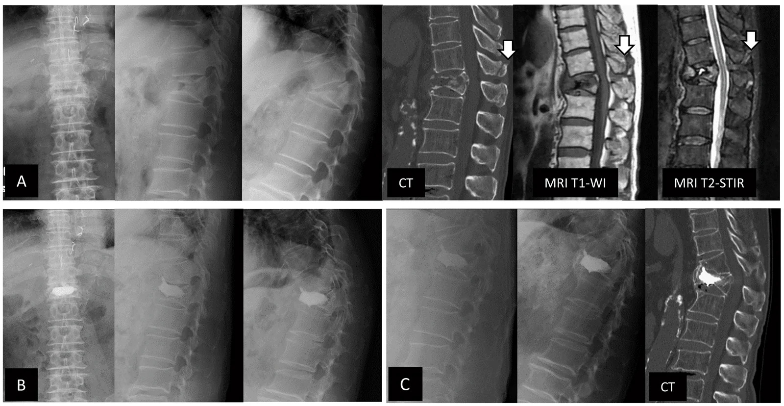
80-year-old man with T12 osteoporotic fracture and posterior element injury: radiographs, CT, T1 and STIR MRI before and after balloon kyphoplasty 54 / 60~3 min · at 55:40CasesCase one is the patient from the first slide, with the ending changed by doing the right things.
Stage one: the examination is benign and the standing films show a 25% wedge at L1 with an intact posterior wall and an old healed T8. The prior films date it. Ask the audience for the classification and whether she needs an MRI.
Stage two: Genant grade 2, AOSpine A1 with the M2 modifier for her bone, OF2, and a TLICS of 1: compression morphology, intact posterior ligamentous complex, intact neurology Vaccaro 2005 Vaccaro 2013. Every trauma system says nonoperative, and they are right today. And, more importantly, she now has two vertebral fractures, a prior wrist fracture and a decade of inhaled steroids. She has severe osteoporosis whatever the DXA will say LeBoff 2022. No red flags and no deficit, so MRI is not required for the immediate decision Hirsch 2018 Expert 2023.
Stage three: the plan is the nonoperative package. Analgesia in layers, mobilisation the same day with physiotherapy, a soft support for confidence and the rigid brace declined on the evidence Kim 2014 Kato 2019; the laboratory screen and a DXA; and a standing radiograph at three weeks Blattert 2018. Ask what happens when she comes back in pain.
Stage four: she has failed conservative care by both criteria, pain and function, and the fracture has progressed to 45% with an upgraded OF type but an intact posterior wall and no cleft. Now the MRI earns its keep: edema confirms the fracture is the pain generator, there is no retropulsion, no ligamentous injury and no malignant features Jung 2003. Ask whether she is a candidate for augmentation.
The image is a comparable open-access case: a T12 osteoporotic fracture with T1 and STIR edema before, and the radiographs and CT after, balloon kyphoplasty.
Stage five: the honest conversation. The sham-controlled trials show a small average benefit Buchbinder 2018; the trials in severe acute pain and the appropriateness pathway support offering it to a patient with an MRI-positive acute fracture, severe pain, functional failure and progression Clark 2016 Hirsch 2018. She chose kyphoplasty, mobilised the same evening, and had pain of 3 out of 10 at two weeks. Her DXA T-score was minus 3.4; with a fresh vertebral fracture she is very high risk, and she started teriparatide with calcium and vitamin D under a fracture liaison service Camacho 2020 Kendler 2018. No new fracture at 18 months.
The point of the case is the sequence: date it, classify it, treat it properly, follow it with standing films, escalate on failure, and treat the bone from day one.
Pearl: Progression on standing films plus failure of pain and function is the trigger to reimage and reconsider, not a reason to add a stronger opioid.An 82-year-old man with a burst fracture
Lateral radiograph: three-level lumbar internal fixator with cement-augmented pedicle screws and cage, 85-year-old woman 55 / 60~2 min · at 58:00CasesCase two is the fracture that must not be called a compression fracture.
Stage one: a rigid kyphotic spine with ankylosing spondylitis, a palpable step, and a new incomplete cord injury: upper motor neuron signs, retention, sacral sparing. Ask the room what the worst possibility is.
Stage two: three things stack up. A neurological deficit; an ankylosed spine, which fractures like a long bone and is unstable; and a delayed deficit two weeks after injury, which means the fracture has moved or collapsed Ito 2002 Blattert 2018. This is not a pain problem.
Stage three: CT shows the truth: a burst-split with a fracture line through the fused anterior and posterior columns, half the canal occupied by a retropulsed fragment, and 30 degrees of focal kyphosis. In OF terms this is OF5; in AOSpine terms a B3 hyperextension injury through an ankylosed spine with an N3 incomplete deficit and the M2 modifier; and in TLICS terms a distraction injury, 4, with a disrupted posterior ligamentous complex, 3, and an incomplete cord injury, 3: ten out of ten, as unambiguous as the score gets Schnake 2018 Vaccaro 2013 Vaccaro 2005. The confounders chapter's warning about the ankylosed spine applies in full: long lever arms, a transverse fracture that hid on the first radiograph, and a low threshold for fixation Williams 2011. MRI shows cord compression with signal change, an epidural haematoma, and edema through the posterior elements. Retropulsion beyond 42% is one of the imaging predictors of neurological impairment in Muratore's review Muratore 2018. The Hounsfield units of 70 tell you the bone is very poor before anyone has ordered a DXA Schreiber 2011.
Stage four: immediate care is spinal precautions with the spine kept in his habitual kyphosis, because forcing an ankylosed spine into extension on a flat board can complete the fracture; a catheter; reversal of anticoagulation in discussion with haematology; and an urgent surgical consultation. The operation is posterior decompression and long-segment cement-augmented fixation, three levels above and below because of the long lever arms of a fused spine and the bone quality, with augmentation of the index level so the anterior column can share load Uchida 2010 Zhang 2021 Blattert 2018.
Stage five: he mobilised on day two, had the pneumonia that these patients get, went to rehabilitation and was walking with a frame and voiding at three months. His bone was treated: vitamin D repletion then zoledronate, with an anabolic declined for practical reasons, and a fracture liaison service to keep him on treatment.
The numbers behind the urgency: in Westerveld's review of ankylosed-spine fractures, two thirds of ankylosing spondylitis patients had a deficit on admission, half had complications, and one in five was dead at three months Westerveld 2009; the deficit often appears days after the injury as the fracture displaces. Timing follows Kerwin's lesson: fix early, within days, once resuscitated and reversed, not on the night of admission in an unoptimised patient Kerwin 2005.
Contrast the two cases: same age group, same "compression fracture" label at first contact, opposite problems. The examination, the posterior wall and the ankylosed spine made the difference.
Pearl: In an ankylosed spine, a 'compression fracture' with a step or a deficit is an unstable long-bone fracture of the spine. Keep it in its own alignment and call the surgeon.- 56 / 60~1 min · at 58:45Cases
The last question, and the one with the largest effect on the patient's future. C. The first fracture is the moment of highest risk and the moment to treat Lindsay 2001 Ebeling 2019. The guidelines put a recent fracture into the very-high-risk category with anabolic-first sequences, and bisphosphonates remain the standard first-line choice where anabolics are unavailable or declined Camacho 2020 Qaseem 2023. Bisphosphonates do not impair vertebral fracture healing, and augmentation, whatever it does for pain, does nothing for the next vertebra.
Refs: Lindsay et al., JAMA, 2001 · Ebeling et al., J Bone Miner Res, 2019 · Camacho et al., Endocr Pract, 2020 · Qaseem et al., Ann Intern Med, 2023 - 57 / 60~1 min · at 59:45Summary
Seven take-home points; this is the slide people photograph.
One: sentinel event. Two: the imaging pathway. Three: the malignancy red flags. Four: classification and the three pivots. Five: nonoperative care as an active package and the brace evidence. Six: augmentation as a selected option, not a reflex. Seven: treat the bone.
If people remember only the first and last points, their patients will have fewer fractures.
- 58 / 60~1 min · at 60:15Summary
Close on the theme. The vertebral fracture is common, under-recognised, sometimes dangerous, and always a warning. Everything in this talk comes back to two decisions: is this the benign kind, and what are we doing about the next one?
Thank the audience and open for questions.
- 59 / 60~0 min · at 60:25Summary
Selected references. The complete list of 92 verified sources with PubMed and DOI links is on the lecture site; the QR code on the next slide takes the audience there.
- 60 / 60~0 min · at 60:40Summary
Leave this slide up during questions. The link opens the deck page with the complete, filterable reference list with PubMed links, the speaker-notes handout, and the research record.