Central Cord Syndrome
Speaker notes · 45 min · v1.0 · updated 2026-09-26
- 1 / 51~1 min · at 0:30Introduction
Welcome. Over the next 45 minutes I want to change how you think about the older patient who falls, hits their forehead, and tells you their hands feel like they are wearing thick gloves.
Central cord syndrome is the most common incomplete spinal cord injury you will see, it is becoming more common as the population ages, and it is missed or under-treated in the emergency department far more often than a fracture-dislocation because the CT is frequently normal.
This talk is built for the people who see these patients first: residents on the consult pager, emergency physicians, and advanced practice providers. Every recommendation is referenced; the small citation chips on the slides can be clicked to show the source, and the full reference list with PubMed links is available online.
- 2 / 51~1 min · at 1:15Introduction
Start with the patient. Paint the picture: a low-energy fall, a forehead laceration or a bruise on the chin, a patient who is ambulatory, perhaps a little unsteady, and who complains that his hands are clumsy, numb or burning. The triage note may say "fall, hand numbness." The CT shows spondylosis and no fracture.
Ask the audience: what happens to this patient in your department if nobody recognises the pattern? He gets a collar clearance, a hand X-ray, and a discharge. That is the failure mode we are trying to prevent.
The forehead or chin injury is the tell: it means the neck was forced into extension. Keep this patient in mind; we will return to him in the cases at the end.
Pearl: A facial or forehead injury in an older patient with new hand symptoms should be treated as a hyperextension cord injury until proven otherwise. - 3 / 51~1 min · at 1:45Introduction
Run through the objectives quickly. Emphasise that objectives four and five are where the evidence has moved most in the last five years: the hemodynamic targets were rewritten in 2024, and the timing-of-surgery literature now includes a propensity-matched central cord cohort and three meta-analyses that did not exist when many of us trained.
Tell the audience explicitly that when a recommendation is opinion rather than evidence, the slide will say so.
- 4 / 51~0 min · at 1:55Why it matters
Section one: the scale of the problem. Two minutes.
- 5 / 51~1 min · at 3:10Why it matters
Three separate lines of evidence tell the same story.
First, among patients admitted to rehabilitation with a defined clinical syndrome, central cord syndrome is the most common, accounting for roughly 44% of the syndromes in McKinley's series, and these were the oldest patients with the lowest admission function scores McKinley 2007.
Second, the demographics of spinal cord injury are shifting. In Thompson's 11-year single-centre series the mean age rose by 13 years and the proportion of injuries that were central cord syndrome rose from a quarter to more than a third Thompson 2015. Jain's analysis of the US Nationwide Inpatient Sample showed that the overall incidence of SCI is stable at about 54 per million, but incidence in men aged 65 to 74 rose sharply, and the proportion of SCI caused by falls in patients over 65 more than doubled to two thirds Jain 2015.
Third, this is a large population: Brodell identified more than 16,000 central cord admissions in eight years of national inpatient data; about 40% had surgery, the surgical rate was rising, and inpatient mortality was 2.6% overall but climbed with age and comorbidity Brodell 2015.
The take-home: this is an older-adult, low-energy, fall-related injury, and the emergency department is where it is either recognised or missed.
- 6 / 51~1 min · at 4:10Why it matters
A brief history, because the controversies in this talk make more sense when you know where they came from.
Richard Schneider and colleagues in Michigan described the syndrome in 1954 in a paper that also proposed the hyperextension mechanism Schneider 1954. His 1958 follow-up consolidated the clinical picture and the tendency to recover legs before arms Schneider 1958. Because he saw good spontaneous recovery, and because operating on these patients in the 1950s carried real risk, the early literature was strongly non-operative. Bosch's ten-year review from Rancho Los Amigos reinforced the idea of a favourable natural history Bosch 1971.
In 1992 Quencer correlated MRI with autopsy specimens and found something important: no blood in the cord, an intact central gray matter, and diffuse axonal disruption in the lateral white matter where the corticospinal tracts run Quencer 1992. This dismantled the textbook idea of a central hematomyelia and we will come back to it.
The modern surgical era started with observational series suggesting that decompression helped, and by 2010 Lenehan's analysis of the Spine Trauma Study Group database was showing a motor-score benefit for surgery within 24 hours in stenotic patients without instability Lenehan 2010.
The 2013 AANS/CNS guidelines and the 2017 AOSpine guidelines codified ICU care, MAP targets and early surgery as an option, with low-quality evidence Aarabi 2013 Fehlings 2017. The 2022 JAMA Surgery propensity-matched cohort was the first central cord specific analysis with modern methods Badhiwala 2022, and the 2024 AO Spine and Praxis update moved early surgery from "an option" to "the preferred option" for all acute SCI, while also loosening the MAP targets Fehlings 2024 Kwon 2024.
- 7 / 51~0 min · at 4:20Anatomy & mechanism
Section two. About six minutes. The goal is that the audience can explain to a patient why their hands are the problem when their legs are fine.
- 8 / 51~1 min · at 5:00Anatomy & mechanism
This is the working definition, essentially unchanged from Schneider's original description and used verbatim in the modern systematic reviews Schneider 1954 Anderson 2015.
Three parts: it is incomplete, so there is sacral sparing; the arms are weaker than the legs, and within the arms the hands are the worst; and there is a variable sensory and bladder component. The typical patient has a narrowed canal from spondylosis and a hyperextension mechanism, and often no bony injury.
Hold on to the phrase "disproportionate weakness." How much disproportion counts is the subject of an entire slide later, because it turns out nobody agrees.
Refs: Schneider et al., J Neurosurg, 1954 · Anderson et al., Neurosurgery, 2015 - 9 / 51~2 min · at 7:00Anatomy & mechanism
Build this diagram in four steps.
Step one: orient the audience. Anterior is up. The gray matter butterfly contains the motor neurons in the anterior horns, including the pools for the hand at C8 and T1. The surrounding white matter carries the long tracts.
Step two: the lateral corticospinal tract sits in the lateral column. This is the primary descending motor pathway, and in primates it is disproportionately devoted to skilled hand and finger movement.
Step three: the classic explanation. For decades we taught that the corticospinal tract is somatotopically laminated, cervical fibres medially and sacral fibres laterally, so that an injury spreading outward from the centre of the cord would hit the arm fibres first and spare the legs. It is an elegant story and it appears in many textbooks. The problem is that the anatomical evidence does not support it. Levi, Tator and Bunge reviewed the primate literature in 1996 and concluded there is no such somatotopic organisation in the medulla or cervical cord Levi 1996. Bucy's observations after deliberate pyramidal tract section in humans had already shown that a corticospinal lesion chiefly impairs fine hand movement rather than walking Bucy 1964.
Step four: what the injury actually looks like. Quencer's MRI-pathology correlation found no hemorrhage, an intact central gray matter, and diffuse axonal disruption in the lateral columns in the region of the corticospinal tracts Quencer 1992. Jimenez and Levi's histopathology series confirmed primary injury to the lateral corticospinal tract with Wallerian degeneration in every case, without loss of the hand motor neurons in acute cases Jimenez 2000.
So the modern explanation for "hands worse than legs" is functional rather than anatomical: the corticospinal tract is essential for hand dexterity but not for locomotion, which relies heavily on other descending and spinal pattern-generating systems. Injure the lateral columns and you lose the hands; the legs walk on the pathways that survived.
Why does this matter clinically? Because it reframes central cord syndrome as a white-matter, compression-related injury of the corticospinal tracts, which is exactly the kind of injury where relieving persistent compression is biologically plausible.
Pearl: The name is a historical accident: the injury is not central. It is a lateral-column corticospinal injury, and hand function is what the corticospinal tract is for.Refs: Levi et al., Neurosurgery, 1996 · Quencer et al., Neuroradiology, 1992 · Jimenez et al., Spinal Cord, 2000 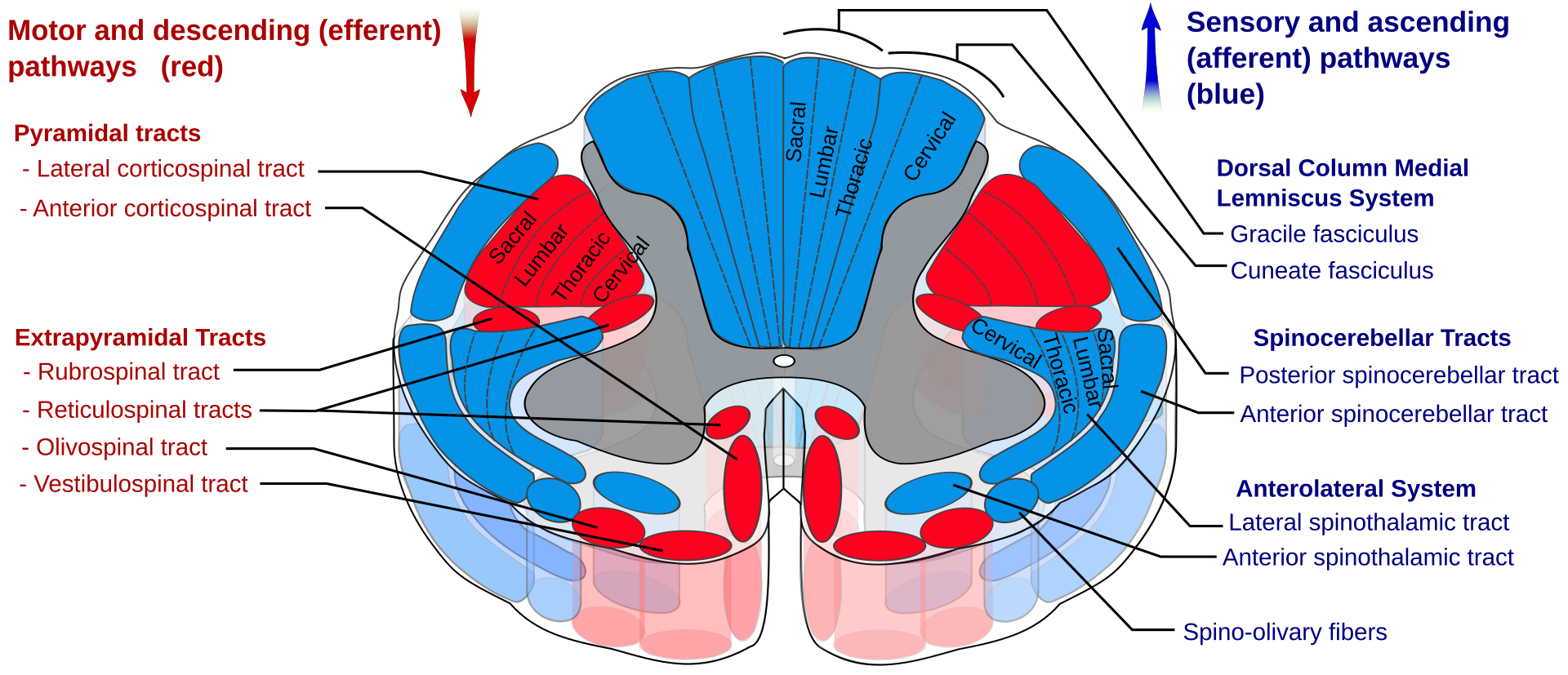
The picture in every textbook
10 / 51~1 min · at 7:40Anatomy & mechanismAsk the room: what is wrong with this picture? Pause.
Nothing is wrong with it as a summary of the tracts. What is wrong is the inference we drew from it. The neat lamination of the lateral corticospinal tract, cervical inside and sacral outside, is drawn in most textbooks and was the basis of the "central lesion spares the legs" story. The primate anatomical evidence does not support that lamination Levi 1996, and the pathology shows the injury is in the lateral columns rather than radiating from the centre Quencer 1992. The honest modern explanation is functional: the corticospinal tract is what the hand depends on.
Diagram by Polarlys and Mikael Häggström, Wikimedia Commons, Creative Commons Attribution-ShareAlike.
- 11 / 51~2 min · at 9:10Anatomy & mechanism
This is Schneider's original mechanism, and the modern imaging data support it Schneider 1954 Quencer 1992.
Stage one: the baseline. An older cervical spine has lost disc height, has anterior disc-osteophyte complexes, and has a thickened, less elastic ligamentum flavum. The sagittal diameter of the canal is already reduced. In Aarabi's surgical series of stenotic central cord patients the mean midsagittal diameter at the point of maximum compression was 5.6 millimetres and the mean canal compromise was about 50% Aarabi 2011.
Stage two: extension. When the neck is forced backward, the canal shortens and the ligamentum flavum buckles forward into the canal. At the same time the anterior osteophytes are driven backward. Quencer suggested that infolding of the ligamenta flava into an already narrow canal is probably the most common mechanism Quencer 1992.
Stage three: the pincer. The cord is squeezed between bone in front and ligament behind. The compression is often transient; the neck returns to neutral, there is no fracture and no dislocation, and the CT shows only degenerative change. That is why the CT report reads "no acute abnormality" in a patient with a cord injury.
Emphasise the clinical corollary for the emergency department: in this population, a normal CT does not exclude a cord injury. A neurological deficit is the indication for MRI, not the CT result.
Also mention the younger mechanism briefly: high-energy hyperextension or flexion injuries with fracture, facet dislocation or acute disc herniation can produce the same clinical syndrome with a structurally unstable spine; we will compare the two on the next slide.
- 12 / 51~1 min · at 10:25Anatomy & mechanism
Central cord syndrome is one clinical picture with two very different structural stories.
The older patient with spondylosis is the majority. In Aarabi's series the mean age was 58, more than half were falls, and the compression was most often at C3-4 and C4-5 Aarabi 2011. The spine is stable. The question for the surgeon is whether persistent cord compression is worth relieving, and when.
The younger patient has a high-energy mechanism and a structural injury: a fracture, a facet dislocation, or an acute traumatic disc herniation. Here the operative decision is driven by instability, and reduction and stabilisation are needed regardless of the cord syndrome. Guest's series of 50 surgically treated patients is the classic illustration: decompression within 24 hours improved motor recovery and shortened ICU and hospital stay in patients with disc herniation or fracture, but not in those with stenosis or spondylosis alone Guest 2002. That distinction still runs through the timing literature today.
Avila and Hurlbert have proposed simply redefining central cord syndrome as any adult cervical cord injury without fracture or dislocation, precisely to separate these two populations for research Avila 2021. Whether or not that catches on, you should mentally sort your patient into one of these two columns before you call the surgeon.
Refs: Guest et al., J Neurosurg, 2002 - 13 / 51~0 min · at 10:35Recognition
Section three, the heart of the talk for emergency clinicians: eight to ten minutes.
- 14 / 51~2 min · at 12:05Recognition
The history is the diagnosis in most cases.
The hands are always the headline complaint. Patients describe weakness of grip and loss of fine motor control: buttons, keys, cutlery. Distal upper limb weakness out of proportion to proximal weakness is characteristic.
Sensory symptoms are typically a burning dysesthesia in the hands and forearms. Merriam's reappraisal of 77 patients noted that hyperpathia and a Lhermitte sign were common and, interestingly, associated with a better prognosis Merriam 1986.
The legs are relatively spared. That is the trap: an ambulatory patient does not look like a spinal cord injury to a busy department. Some have subtle spasticity or unsteadiness; ask them to walk heel-to-toe.
Bladder dysfunction is variable. Urinary retention is the usual pattern and is part of most definitions, so a post-void residual in the department is worthwhile; it also matters for prognosis and for early catheter management.
Sacral sparing is what makes the injury incomplete. You must examine for it: perianal pinprick and light touch at S4-5, deep anal pressure, and voluntary anal contraction. Without this examination you cannot assign an AIS grade.
Finally, the mechanism clue. Neck pain can be mild. Look at the face: a laceration on the forehead, a bruise on the chin, a broken nose. These tell you the neck was extended.
A brief differential, because most of the work is pattern recognition. The other incomplete syndromes: anterior cord syndrome gives motor loss with loss of pain and temperature and preserved proprioception; Brown-Séquard is a hemicord picture; cruciate paralysis at the cervicomedullary junction produces the same arm-worse-than-leg pattern and probably shares the corticospinal explanation Levi 1996. Chronic cervical spondylotic myelopathy is often unmasked by a fall; months of clumsy hands and unsteady gait before the injury change the counselling but not the acute workup. A bilateral brachial plexus injury after a fall onto both shoulders has absent rather than brisk reflexes, patchy sensory loss and a normal sacral examination. Head injury coexists often enough to be suspicious of both, and a depressed level of consciousness makes the cord examination unreliable and pushes you toward MRI. And always look again at the CT for facet subluxation, a widened interspinous space, a traumatic disc, or a fracture through an ossified ligament or an ankylosed spine.
- 15 / 51~1 min · at 13:05Recognition
Quantify the disproportion, because the literature does.
The ISNCSCI examination scores five key muscles in each limb on the 0 to 5 Medical Research Council scale. Summing both sides gives an upper extremity motor score and a lower extremity motor score, each out of 50 Rupp 2021.
In Aarabi's surgical series of stenotic central cord patients the admission means were 25.8 for the arms and 39.8 for the legs Aarabi 2011. That 14-point gap is a typical picture.
Pouw's systematic review of 312 patients from nine studies found that the mean upper extremity score was 10.5 points below the lower extremity score, and proposed 10 points as a diagnostic criterion Pouw 2010. When van Middendorp surveyed 157 AOSpine surgeons, definitions varied widely but 61% accepted a 10-point difference van 2010.
Note that the JAMA Surgery propensity-matched study used a five-point threshold Badhiwala 2022, and Engel-Haber's 2023 review lists several competing definitions and argues that none captures the whole syndrome Engel-Haber 2023. So when you read a paper, check how the authors defined their cohort.
Pearl: Write the two numbers in your note: UEMS and LEMS. It is the fastest way to communicate the syndrome to the surgeon and to track recovery.Refs: Rupp et al., Top Spinal Cord Inj Rehabil, 2021 · Aarabi et al., J Neurosurg Spine, 2011 · Pouw et al., Spinal Cord, 2010 · van et al., Spinal Cord, 2010 - 16 / 51~1 min · at 14:20Recognition
Five competing definitions in one table. You do not need to memorise them; you need to know that they exist, because they explain why two papers on "central cord syndrome" can reach different conclusions.
Schneider's description is the one everyone learns and it remains the clinical gestalt Schneider 1954. The 10-point motor difference from Pouw's systematic review is the most widely used research criterion Pouw 2010. The JAMA Surgery propensity-matched study, which we will discuss in the timing section, used 5 points Badhiwala 2022. Avila and Hurlbert argued for a structural definition, any adult cervical cord injury without fracture or dislocation, on the grounds that it would make the surgical literature comparable Avila 2021. And there are two published scoring systems that add MRI signal and functional items for prognosis Hohl 2010 Ramey 2021.
The practical message comes from the prospective European cohort: among 248 motor-incomplete tetraplegics, the AIS grade was the best prognostic parameter and functional outcomes did not differ according to the size of the upper-lower limb gap Pouw 2011. In other words, the label matters less than the severity. Document the AIS grade and the motor scores and you have captured what matters.
Refs: Schneider et al., J Neurosurg, 1954 · Pouw et al., Spinal Cord, 2010 · Badhiwala et al., JAMA Surg, 2022 · Avila & Hurlbert, Neurosurg Clin N Am, 2021 · Hohl et al., Spine (Phila Pa 1976), 2010 · Ramey et al., World Neurosurg, 2021 · Pouw et al., Spinal Cord, 2011 · Engel-Haber et al., Spinal Cord, 2023 - 17 / 51~2 min · at 16:00Recognition
The International Standards for Neurological Classification of Spinal Cord Injury, the ASIA examination, is the language of this field. The current version is the 2019 revision Rupp 2021; the 2011 revision defined the scoring most of the literature uses Kirshblum 2011. The worksheet is freely downloadable from ASIA and I encourage every department to keep it in the trauma bay.
Motor: ten key muscles on each side, five in the arm and five in the leg, graded 0 to 5. The arm muscles are elbow flexors C5, wrist extensors C6, elbow extensors C7, finger flexors C8 and finger abductors T1. The leg muscles are hip flexors L2, knee extensors L3, ankle dorsiflexors L4, long toe extensors L5 and ankle plantar flexors S1. Sum them: upper extremity motor score and lower extremity motor score, 50 each.
Sensory: light touch and pinprick in 28 dermatomes per side, scored 0 to 2, and this must include S4-5.
The sacral examination is non-negotiable: perianal sensation, deep anal pressure and voluntary anal contraction. It is what defines an incomplete injury, and therefore whether the patient is AIS B, C or D rather than A.
Then determine the neurological level and the AIS grade and write down the time of the examination. Every prognostic study finds that the admission motor score is the strongest predictor of the final score Aarabi 2011 Dvorak 2005 Aarabi 2021, and the AIS grade at presentation is the variable on which the surgical timing evidence turns: in the JAMA Surgery cohort, early surgery benefited AIS C patients substantially and AIS D patients not at all Badhiwala 2022.
One caution: the first examination may be affected by pain, alcohol, sedation, head injury and spinal shock. Ditunno's four-phase model reminds us that reflexes evolve over days Ditunno 2004. Re-examine at 24 and 72 hours. Trends matter more than a single snapshot.
Refs: Rupp et al., Top Spinal Cord Inj Rehabil, 2021 · Kirshblum et al., J Spinal Cord Med, 2011 · Aarabi et al., J Neurosurg Spine, 2011 · Dvorak et al., Spine (Phila Pa 1976), 2005 · Aarabi et al., J Neurotrauma, 2021 · Lenehan et al., Spine (Phila Pa 1976), 2010 · Badhiwala et al., JAMA Surg, 2022 · Ditunno et al., Spinal Cord, 2004 - 18 / 51~1 min · at 16:45Recognition
Walk down the ladder.
AIS A is complete: no motor or sensory function in the lowest sacral segments. B is sensory incomplete: sensation preserved at S4-5 but no motor function more than three levels below the motor level. C and D are motor incomplete, and the dividing line is whether at least half of the key muscles below the neurological level have a grade of 3 or better. E is normal in a patient who had a documented prior deficit Rupp 2021.
Central cord patients are overwhelmingly C or D. The distinction between C and D is not academic. In Lenehan's analysis the expert consensus was that early decompression was reasonable for AIS C patients with persistent compression, whereas AIS D patients could be observed initially Lenehan 2010. In the 2022 propensity-matched cohort, the benefit of early surgery was concentrated in AIS C patients, who gained about 9.5 motor points, with no difference in AIS D Badhiwala 2022.
So the grade you assign in the department feeds directly into the surgical decision.
Refs: Rupp et al., Top Spinal Cord Inj Rehabil, 2021 - 19 / 51~1 min · at 17:45Recognition
Two terms that sound alike and are constantly confused.
Spinal shock is a neurological phenomenon: the temporary loss of reflexes and tone below the injury. Ditunno's four-phase model describes areflexia in the first day, initial reflex return, and then early and late hyperreflexia over weeks Ditunno 2004. Its clinical relevance for us is that a patient in spinal shock may look complete when they are not, so the AIS grade should be re-checked as reflexes return. It does not cause hypotension.
Neurogenic shock is hemodynamic: hypotension and often bradycardia caused by loss of sympathetic tone below a cervical or high thoracic injury. Ruiz and colleagues showed that the reported incidence depends entirely on the definition, with a figure around 29% in cervical SCI when combined hemodynamic and laboratory criteria were used, and that hypovolemia is a major confounder Ruiz 2018.
In the trauma bay this means: assume hemorrhage until excluded, give volume, and then use a vasopressor. Because the problem is loss of sympathetic tone with bradycardia, an agent with both alpha and beta activity, such as norepinephrine, is the usual choice; pure alpha agonists can worsen bradycardia. We will come to specific pressor data in older central cord patients in the management section Readdy 2015.
Central cord patients are less likely to have profound neurogenic shock than complete cervical injuries, but hypotension in this group is common for other reasons: age, medications, dehydration and blood loss from a facial injury. Whatever the cause, hypotension is bad for the injured cord.
Refs: Ditunno et al., Spinal Cord, 2004 · Ruiz et al., J Neurotrauma, 2018 - 20 / 51~1 min · at 18:45Recognition
Give the audience 20 seconds. Then reveal.
The correct answer is MRI and a spine consultation now. The teaching points: the decision rules for imaging apply to patients without a neurological deficit; a negative CT excludes most fractures but not a cord injury in a stenotic canal; and MRI is the study that shows the cord, the compression and the ligaments Hoffman 2000 Ryken 2013 Bozzo 2011.
Flexion-extension films in an acutely injured patient with a deficit are both unhelpful and potentially harmful. Nerve conduction studies answer a different question on a different timescale.
This leads us straight into the imaging section.
Refs: Hoffman et al., N Engl J Med, 2000 · Ryken et al., Neurosurgery, 2013 · Bozzo et al., J Neurotrauma, 2011 - 21 / 51~0 min · at 18:55Imaging
Section four: imaging, about six minutes.
- 22 / 51~2 min · at 20:35Imaging
Everything on this slide is guideline-level, so it is worth being precise.
Start with the decision rules. In the alert patient without a deficit, NEXUS and the Canadian C-Spine Rule identify who needs imaging at all. NEXUS was validated in 34,000 patients with 99.6% sensitivity Hoffman 2000; the Canadian rule was derived in almost 9,000 alert, stable patients with 100% sensitivity for clinically important injury Stiell 2001. Both rules require a normal neurological examination. A patient with weak hands has failed the rule by definition.
When imaging is indicated, the study is CT. The EAST guideline replaced plain films with CT as the primary modality in 2009 Como 2009, based largely on Holmes's meta-analysis showing pooled sensitivity of 98% for CT versus 52% for radiographs Holmes 2005. The 2013 AANS/CNS radiographic assessment guideline says the same Ryken 2013.
Now the deficit branch, which is the point of this talk. The patient with a neurological deficit gets a CT, because you need to know about fracture, dislocation, OPLL and canal size, and then gets an MRI regardless of the CT result. The MRI shows the cord signal change, persistent compression, disc herniation and ligamentous injury; Bozzo's review and Delphi vote strongly recommended MRI for prognostication in acute cord injury Bozzo 2011. Schoenfeld's meta-analysis found that even in patients with a negative CT, MRI found abnormalities in 12% and changed management in 6% Schoenfeld 2010.
The obtunded patient is a special case. The 2015 EAST guideline conditionally recommends removing the collar after a negative high-quality CT Patel 2015. But an obtunded patient cannot give you an examination, so if there is any clinical suggestion of a cord injury, for instance an unexplained flaccid limb or priapism, or if the CT is equivocal, get the MRI.
And the footnote: flexion-extension films have no place in the acute injured patient with a deficit.
Refs: Hoffman et al., N Engl J Med, 2000 · Stiell et al., JAMA, 2001 · Como et al., J Trauma, 2009 · Holmes & Akkinepalli, J Trauma, 2005 · Bozzo et al., J Neurotrauma, 2011 · Ryken et al., Neurosurgery, 2013 · Patel et al., J Trauma Acute Care Surg, 2015 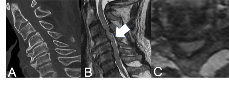
Normal CT. Abnormal cord.
23 / 51~1 min · at 21:15ImagingThis is the picture that should be burned into memory. On the left, a sagittal CT with degenerative change and no fracture, the kind of study that gets read as "no acute abnormality." In the middle, the T2 MRI from the same patient with a bright intramedullary signal at C3/4: cord edema at the level of maximal narrowing. On the right, the axial view.
The CT is not wrong. It answers the question it was asked, which is whether there is a fracture. It cannot see the cord. This is why the examination, not the CT, decides whether an MRI is needed.
Image from an open-access case report, reproduced under a Creative Commons licence with attribution shown on the slide.
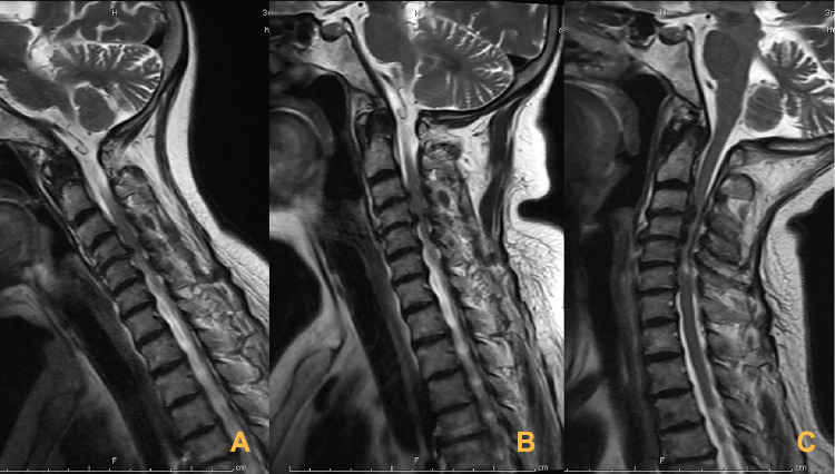
The mechanism, caught on MRI
24 / 51~1 min · at 21:55ImagingKinematic MRI makes Schneider's mechanism visible. The same patient is scanned in flexion, neutral and extension. In extension the anterior disc-osteophyte complexes and the infolded ligamentum flavum converge on the cord. In neutral, which is how the patient reaches your department, the compression looks far less dramatic.
Two lessons. First, the static neutral MRI underestimates what happened at the moment of injury. Second, this is why we keep these patients in a collar and avoid extension during airway management and transfers until the surgeon has made a plan.
Image reproduced from Li and colleagues, Therapeutics and Clinical Risk Management 2021, under a Creative Commons non-commercial licence.
What matters on the MRI
- T2 cord signal: edema is expected; hemorrhage on T2* or T1 predicts worse recovery Bozzo 2011 Wilson 2012
- Length of intramedullary hyperintensity: longer lesions predict worse motor outcome and pain Aarabi 2011 Aarabi 2021
- Degree of compression: midsagittal diameter and maximum canal compromise predict outcome Aarabi 2011
- Persistent compression after injury is the surgical target; document its level and cause
- STIR/fat-saturated sequences: ligamentous injury, interspinous edema, disc disruption
- The BASIC score grades axial T2 involvement 0–4 and tracks with AIS grade Talbott 2015
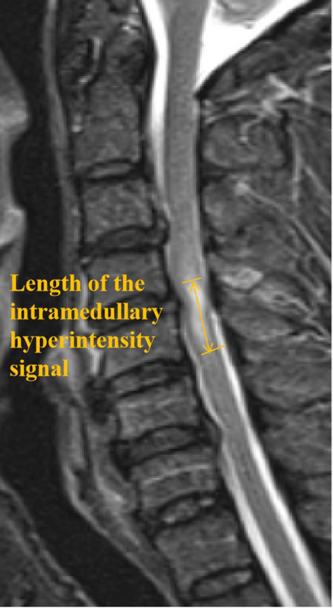
Lesion length measured on sagittal T2. 25 / 51~2 min · at 23:35ImagingWhat should you read off the MRI, and what should you ask the radiologist to comment on?
Cord signal. T2 hyperintensity, edema, is expected in central cord syndrome and by itself is not alarming. Hemorrhage within the cord is a different matter: in Bozzo's synthesis of sagittal T2 patterns, and in Wilson's prediction model, hemorrhage and multilevel edema predicted worse outcome Bozzo 2011 Wilson 2012. Remember from Quencer that hemorrhage is uncommon in true central cord syndrome Quencer 1992, so its presence suggests a more severe injury.
Lesion length. In Aarabi's series the mean length of parenchymal damage was about 29 millimetres, and lesion length predicted manual dexterity and dysesthetic pain at one year Aarabi 2011. In the 2021 cohort intramedullary lesion length was 23 millimetres on average and, along with admission motor score, was the variable that mattered Aarabi 2021.
Compression. Midsagittal diameter at the tightest level and maximum canal compromise both predicted outcome Aarabi 2011. Persistent compression after the injury is the thing the surgeon can change, so note the level and whether it is from disc, osteophyte, OPLL or ligament.
Ligamentous injury. Fat-saturated T2 or STIR shows interspinous edema and disruption of the posterior ligamentous complex, which changes a stable-looking spine into an unstable one and is a component of the SLIC and AOSpine classifications Vaccaro 2007 Vaccaro 2016.
Finally, for those who want a reproducible score, the BASIC score from UCSF grades the axial T2 pattern from 0 to 4 and correlates with AIS grade at admission and discharge Talbott 2015.
Refs: Bozzo et al., J Neurotrauma, 2011 · Wilson et al., J Neurotrauma, 2012 · Aarabi et al., J Neurosurg Spine, 2011 · Aarabi et al., J Neurotrauma, 2021 · Talbott et al., J Neurosurg Spine, 2015What the CT can add
- Spondylotic stenosis without fracture: the majority in older adults
- OPLL: ossified posterior longitudinal ligament, common in East Asian populations, may fracture through the ossified mass
- Traumatic disc herniation: young patient, normal bone, acute cord compression
- Fracture or facet dislocation: unstable; reduce and stabilise
- Ankylosed spine (DISH, ankylosing spondylitis): extension fractures through a fused column are unstable and easily missed
- Classify structural injuries with SLIC or the AOSpine subaxial system to frame the operative decision Vaccaro 2007 Vaccaro 2016
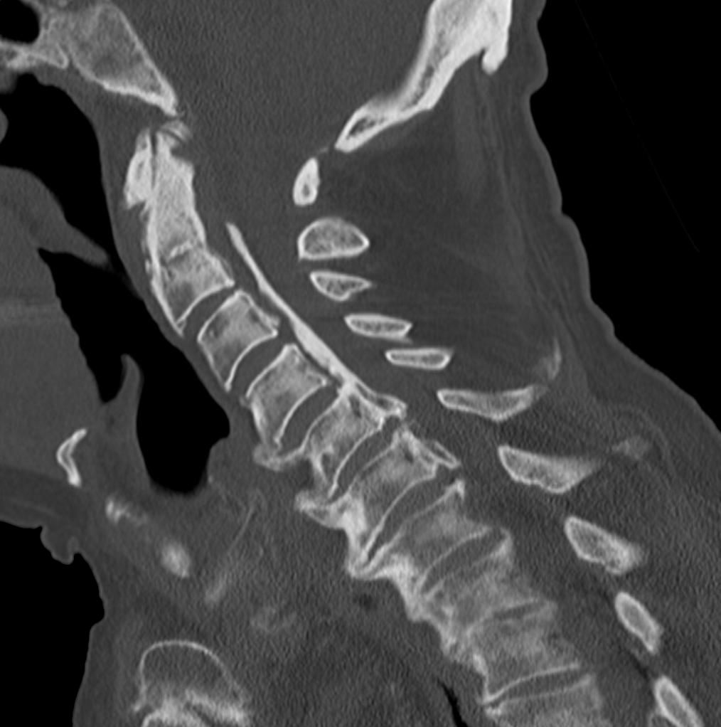
Sagittal CT: ossification of the posterior longitudinal ligament. 26 / 51~1 min · at 24:50ImagingThe CT still has a lot to say, even when it shows no fracture.
Most older patients have multilevel spondylotic stenosis. Measure or at least eyeball the canal; a developmentally narrow canal makes the whole cord vulnerable.
OPLL, ossification of the posterior longitudinal ligament, is shown in the aside. It is much more common in East Asian populations and it matters because a rigid ossified mass in front of the cord both narrows the canal and, after hyperextension, can fracture, creating an unstable segment that is easy to miss.
Traumatic disc herniation is the young person's version: normal bone on CT, acute soft disc compression on MRI, and a surgical target.
Fractures and facet dislocations are unstable and the treatment is reduction and stabilisation, whatever the cord syndrome.
The ankylosed spine deserves special mention: in DISH or ankylosing spondylitis, a low-energy extension injury can fracture through the fused column, the fracture is often subtle on CT, and the spine is grossly unstable. These patients are frequently misdiagnosed as having a simple central cord syndrome. If the spine is fused, look for the fracture until you find it, and keep the patient flat and immobilised.
Finally, the SLIC and AOSpine subaxial classifications score morphology, discoligamentous integrity and neurology, and are the common language between you and the surgeon for structural injuries Vaccaro 2007 Vaccaro 2016.
Refs: Vaccaro et al., Spine (Phila Pa 1976), 2007 · Vaccaro et al., Eur Spine J, 2016- 27 / 51~0 min · at 25:00Initial management
Section five, initial management in the emergency department and the first 24 hours. About eight minutes, including the steroid evidence.
- 28 / 51~2 min · at 26:40Initial management
A pathway for the first hours. Most of it is standard trauma care applied with the cord in mind.
Spinal motion restriction: a collar, log-rolling and, above all, no extension of the neck. Airway manoeuvres in this population should be done with in-line stabilisation and a video laryngoscope or fibreoptic technique where available.
Airway and breathing. Central cord patients usually breathe adequately, but a high injury, edema ascending over the first days, and aspiration in an older patient lying flat can all cause deterioration. Respiratory failure and pneumonia were the leading complications in Grossman's prospective cohort Grossman 2012, and respiratory failure was the leading cause of death in the Welsh five-year cohort Stanley 2023. Watch the vital capacity, and intubate early and in a controlled fashion rather than late in a crisis.
Circulation. Hypotension is bad for the injured cord. First exclude hemorrhage; then treat neurogenic hypotension with volume and a vasopressor. Avoid a systolic pressure below 90 Ryken 2013. We will discuss the MAP target on the next slide.
Examination. This is the moment to do the full ISNCSCI examination properly and write down the time; it is the baseline for every decision that follows.
Imaging as per the previous section.
And then: consult at the time of diagnosis, not the next morning. The evidence on timing of surgery makes this matter. Admit to an ICU or monitored bed; place a urinary catheter, because retention is common and bladder distension can provoke autonomic instability; keep the patient fasted until the plan is clear; start VTE prophylaxis within 72 hours per the 2013 guideline Dhall 2013; and think about pressure areas from the first hour. If the patient is on anticoagulants, hold them and discuss reversal with the surgeon.
Refs: Ryken et al., Neurosurgery, 2013 · Kwon et al., Global Spine J, 2024 - 29 / 51~2 min · at 28:20Initial management
Hemodynamic management is one of the only medical interventions thought to improve neurological outcome, and the target has changed.
The 2013 AANS/CNS guideline recommended maintaining MAP between 85 and 90 mmHg for seven days after injury, as a Level III recommendation, and the central cord chapter repeated it Ryken 2013 Aarabi 2013. Hawryluk's analysis of nearly a million minute-by-minute readings from 100 patients found that higher MAP correlated with recovery in the first two to three days, and that the proportion of time spent below 85 was a stronger correlate than the average, suggesting that duration of hypotension matters most. The authors were explicit that this is correlation, not proof of causation Hawryluk 2015.
Stage two: the 2024 AO Spine and Praxis guideline reviewed the literature and loosened the target. It suggests augmenting MAP to at least 75 to 80 mmHg as a lower limit, not actively pushing beyond 90 to 95, for three to seven days. The quality of evidence was rated very low and the recommendation is weak. The group declined to recommend a specific vasopressor or a spinal cord perfusion pressure target because the evidence is too thin Kwon 2024. The accompanying systematic review found that the effect of MAP support on recovery is uncertain, though higher spinal cord perfusion pressure may be associated with better recovery Evaniew 2024 Squair 2017.
Stage three: the caveats that matter for our older central cord patients. Vasopressors have consequences. In Readdy's series of 34 central cord patients at UCSF, mean age 62, dopamine caused cardiac complications in 68% versus 46% for phenylephrine, and in patients over 55 the difference was significant, 83% versus 50% Readdy 2015. Inoue's larger SCI cohort found dopamine independently associated with complications with an odds ratio near 9 Inoue 2014. So: avoid hypotension, aim for the guideline range, prefer norepinephrine or phenylephrine over dopamine in older patients, use an arterial line, and stop when the patient is clearly recovering rather than pushing an 80-year-old heart for a week.
Pearl: The 2024 target is a floor of 75–80 mmHg for 3–7 days, not a ceiling of 90 for a week. Duration of hypotension probably matters more than the average number.Refs: Ryken et al., Neurosurgery, 2013 · Kwon et al., Global Spine J, 2024 · Hawryluk et al., J Neurotrauma, 2015 · Readdy et al., J Neurosurg Spine, 2015 - 30 / 51~3 min · at 30:50Initial management
Steroids. You will still be asked. Here is the evidence, study by study.
NASCIS II randomised 487 patients to methylprednisolone as a 30 mg/kg bolus followed by 5.4 mg/kg/hour for 23 hours, naloxone, or placebo. The primary analysis showed no difference. A post-hoc analysis of patients treated within eight hours showed a modest improvement in motor scores at six months, a change score of 16 versus 11 Bracken 1990. That subgroup result became the basis for two decades of practice.
NASCIS III compared 24 and 48 hours of methylprednisolone with tirilazad, with no placebo arm, and reported that the 48-hour regimen improved motor recovery if started between three and eight hours, at the cost of more severe sepsis and pneumonia Bracken 1997.
The Cochrane review, written by the NASCIS lead investigator, concluded that high-dose methylprednisolone within eight hours modestly improves motor recovery Bracken 2012.
Evaniew's 2016 meta-analysis applied GRADE methodology to four trials and 17 observational studies and found no long-term motor benefit, a short-term motor gain within eight hours of low confidence, and increased gastrointestinal bleeding in observational data Evaniew 2016.
The guidelines diverged. The 2013 AANS/CNS guideline issued a Level I recommendation against methylprednisolone, citing harm without proven benefit Hurlbert 2013; that recommendation was itself criticised for over-reading the harm data. The 2017 AOSpine guideline took a middle path: it suggests not offering steroids after eight hours, suggests the 24-hour infusion may be offered as an option within eight hours, and suggests never giving the 48-hour regimen Fehlings 2017.
What the evidence shows: a small, uncertain, early-window benefit and real harms. How this affects decisions for our patient population: the typical central cord patient is older, often diabetic, frequently presents late, and is at higher risk of the infectious and gastrointestinal complications. In practice steroids are not a standard of care for central cord syndrome. Do not give them after eight hours. Within eight hours, follow your institution's protocol and the surgeon's preference. If they are used, it is the 24-hour regimen. Whatever you do, document the reasoning.
- 31 / 51~1 min · at 31:35Initial management
A checklist. None of this is glamorous and all of it changes outcomes, because the complications of cord injury are what kill and disable these patients.
Bladder: place a catheter early. Retention is part of the syndrome and an overdistended bladder in a patient with a cervical injury can provoke a hypertensive or bradycardic response.
Respiratory: central cord patients usually breathe well at first, but cord edema can ascend over two to three days and an older patient lying flat in a collar aspirates easily. Measure vital capacity serially.
VTE: the 2013 guideline recommends prophylaxis with low molecular weight heparin or heparin plus mechanical devices, beginning within 72 hours Dhall 2013. In practice: mechanical devices immediately, pharmacological prophylaxis as soon as the surgeon is comfortable.
Skin, temperature, glucose: routine, but they slip when the focus is on the neurological examination.
Serial neurological checks are the safety net. A declining examination is an emergency and the reason the surgeon wants to know early.
Finally, pain. The dysesthetic hand pain can be severe. Treat it, but avoid a level of sedation that masks a change in the examination.
Refs: Dhall et al., Neurosurgery, 2013 - 32 / 51~1 min · at 32:20Initial management
A structured handover makes the surgical decision faster and better.
Time of injury first. Every timing study measures from injury to decompression, and the 2024 guideline threshold is 24 hours from injury Fehlings 2024. If the fall was at 6 pm yesterday, the window is already half gone when the patient arrives at 6 am.
The examination in the surgeon's language: AIS grade, the two motor scores, sacral sparing, bladder. And the trajectory: an improving AIS D patient and a deteriorating AIS C patient are completely different conversations.
The CT findings that define stability, then the MRI findings that define the surgical target.
The physiology, because a patient on two pressors with an unsecured airway is not going to the operating theatre in the next hour whatever the cord looks like.
And the patient: age, comorbidities, anticoagulation, and what they were doing before the fall. An 88-year-old from a nursing home with dementia and an AIS D injury and a 62-year-old golfer with an AIS C injury are managed differently, and the family conversation starts in your department.
- 33 / 51~0 min · at 32:30Surgery & timing
Section six, the controversy. About ten minutes. This is where the literature has moved most, so the evidence slide gets real time.
- 34 / 51~2 min · at 34:00Surgery & timing
First the binary question: operate or not. Then the harder question: when.
Nonoperative management remains reasonable for a specific patient: a stable spine, a milder deficit, especially AIS D, an examination that is improving day by day, little residual compression, or a patient whose medical condition or goals of care make surgery inappropriate. Lenehan's expert consensus stated exactly this: AIS D patients can be observed initially, with surgery later depending on the pattern of recovery Lenehan 2010. Nonoperative management is an active process: serial examinations, MAP support, rehabilitation, and a readiness to change course.
Surgery is indicated when the spine is unstable, and favoured when there is persistent compression with a significant deficit, especially AIS C, when the patient deteriorates or plateaus, and when there is an acute disc herniation, which is the group that gained most from early surgery in Guest's series Guest 2002. Even in patients who recover well, persistent severe stenosis is often decompressed electively to prevent a second injury and progressive myelopathy.
The footnote is important. The 2013 AANS/CNS chapter and the 2019 Spinal Cord Society position statement are consensus documents built on observational data Aarabi 2013 Yelamarthy 2019. There is no randomised trial. A PubMed search performed for this lecture in September 2026 found none.
Refs: Lenehan et al., Spine (Phila Pa 1976), 2010 · Badhiwala et al., JAMA Surg, 2022 · Guest et al., J Neurosurg, 2002 · Aarabi et al., Neurosurgery, 2013 · Yelamarthy et al., Eur Spine J, 2019 - 35 / 51~4 min · at 37:30Surgery & timing
This is the evidence slide. Take it slowly; it is the reason the surgeon wants the call at 2 am.
STASCIS, the Surgical Timing in Acute Spinal Cord Injury Study, enrolled 313 patients with cervical SCI in a prospective multicentre cohort. Those decompressed before 24 hours had a mean time to surgery of 14 hours; the late group, 48 hours. At six months, 19.8% of early patients improved by two or more AIS grades versus 8.8% of late patients, an adjusted odds ratio of 2.8, with no increase in complications Fehlings 2012. It was not randomised, and only 71% had six-month follow-up, but it changed practice.
Lenehan's analysis, specifically in central cord patients with spondylosis and no instability, showed that surgery within 24 hours produced a 6.3-point greater improvement in total motor score at one year and higher odds of AIS improvement Lenehan 2010.
Badhiwala's pooled analysis of individual patient data from four prospective datasets, 1,548 patients, found a 4-point motor score advantage for early surgery at one year, better AIS grades, and, when time was modelled continuously, a steep decline in recovery over the first 24 to 36 hours followed by a plateau Badhiwala 2021. That curve is the next slide.
Then the central cord specific study. Badhiwala 2022 in JAMA Surgery propensity-matched 93 early and 93 late central cord patients from three prospective datasets. Early surgery improved upper limb motor recovery by 2.3 points, not lower limb, and in the pre-planned subgroup analysis AIS C patients gained 9.5 total motor points with early surgery while AIS D patients showed no difference Badhiwala 2022. Note the limitations: mean age 48, so younger than the typical stenotic patient, and central cord syndrome defined as a five-point rather than ten-point gap. Okonkwo's accompanying editorial argued that central cord syndrome is likewise a surgical emergency Okonkwo 2022.
Now the counter-evidence, which deserves equal weight. Aarabi's homogeneous cohort of 101 AIS C and D patients with stenotic central cord syndrome and MRI-confirmed decompression found no effect of timing, whether analysed in categories or continuously; only the admission motor score predicted the outcome Aarabi 2021. His earlier analysis found the same for ultra-early surgery in a broader cervical SCI group Aarabi 2020. Kepler's series likewise found no early difference Kepler 2015. In older stenotic patients with good baseline function, the benefit of rushing may be small.
Finally, three meta-analyses appeared in 2024. Sattari pooled nine studies and 5,619 patients: early surgery gave a 4.9-point motor advantage at one year with moderate certainty, fewer venous thromboembolic events and complications, a shorter stay, and no difference in mortality Sattari 2024. Xu's analysis of 13 studies found a 7-point advantage and fewer complications Xu 2024; Bin-Alamer's smaller analysis found fewer surgical complications and a non-significant motor trend Bin-Alamer 2024.
Read the summary boxes aloud. What the evidence shows: moderate-quality observational evidence for early surgery across SCI, a smaller and less consistent signal in central cord syndrome, concentrated in AIS C patients and those with structural compression, and some stenotic cohorts with no timing effect at all. How this affects decision-making: treat persistent compression with a meaningful deficit as time-sensitive, get the surgeon involved at diagnosis, resuscitate fast, and aim for surgery within 24 hours when the patient can tolerate it. AIS D with an improving examination can be watched.
- 36 / 51~1 min · at 38:15Surgery & timing
A picture of the pooled-analysis finding. When time to decompression was modelled as a continuous variable, the expected motor recovery fell steeply over the first 24 to 36 hours and then flattened Badhiwala 2021. Two implications: there is no cliff at exactly 24 hours, so a patient at 26 hours still benefits from getting to theatre promptly; and the biggest gains are in the first hours, which is why the "time is spine" framing from the translational literature has taken hold Badhiwala 2019.
Be honest about the limits: this curve is illustrative, drawn to match the published description, and the underlying data are observational. The 2024 guideline group explicitly declined to recommend ultra-early surgery, within 12 hours, because the studies were small and inconsistent Fehlings 2024 Fehlings 2024.
Refs: Badhiwala et al., Lancet Neurol, 2021 · Badhiwala et al., J Neurosurg Spine, 2019 - 37 / 51~1 min · at 39:30Surgery & timing
Four guideline statements, in order.
The 2013 AANS/CNS chapter on central cord syndrome made Level III recommendations for ICU care, MAP support and early decompression, particularly of a compressed cord with a fracture or dislocation Aarabi 2013.
The 2017 AOSpine guideline, based on a systematic review that found one central cord specific study with a marginal benefit, produced a weak suggestion that early surgery be considered as an option in central cord syndrome, with low-quality evidence Fehlings 2017 Wilson 2017.
The 2019 Spinal Cord Society and Spine Trauma Study Group position statement, built on a systematic review, favoured early surgery in selected patients with persistent compression Yelamarthy 2019.
The 2024 AO Spine and Praxis update is the current standard. On moderate-quality evidence, with a relative risk of 2.76 for a two-grade AIS improvement at six months and 4.5 additional motor points, the group recommended that early surgery within 24 hours be offered as the preferred option for adults with acute SCI regardless of level, and stated that a recommendation on ultra-early surgery could not be made Fehlings 2024 Fehlings 2024.
Notice the language shift over eleven years, from "considered as an option" to "the preferred option." Also notice the 2024 document does not make a separate rule for central cord syndrome; the central cord data were part of the whole. That is a fair reading, but it means the specific counter-evidence in older stenotic patients is not fully resolved.
Practice lags the guidelines. In the ACS Trauma Quality Improvement Program data, 3,928 central cord patients across 255 centres, 39% were treated nonoperatively, and after adjusting for case mix the median odds ratio between centres was 2, meaning the hospital you land in strongly predicts whether you have surgery Badhiwala 2024. In the PearlDiver insurance database only 17% of 11,653 patients with central cord syndrome and no fracture had surgery, though the proportion nearly doubled from 2015 to 2020 Dhodapkar 2024, and among operated patients in the National Inpatient Sample only 17% had surgery within 24 hours of admission, a fraction that is rising Gagliardi 2025. The counter-argument comes from Samuel's National Trauma Data Bank analysis of 1,060 operated patients, in which each 24-hour delay was associated with 19% lower odds of inpatient mortality, most plausibly because sicker patients were rushed to theatre and because medical optimisation in a frail older patient has real value Samuel 2015. The 2024 meta-analysis found no mortality difference either way Sattari 2024. The synthesis: early surgery is the goal for the patient who can tolerate it; in the frail older patient the first 24 hours may be better spent on resuscitation and optimisation, and that is a bedside judgment, not a protocol.
Refs: Aarabi et al., Neurosurgery, 2013 · Fehlings et al., Global Spine J, 2017 · Yelamarthy et al., Eur Spine J, 2019 · Fehlings et al., Global Spine J, 2024 Choosing the operation
- Match the approach to the compression: anterior for one- or two-level disc-osteophyte disease, posterior for multilevel stenosis or OPLL with preserved lordosis
- ACDF is the most common procedure in national data Brodell 2015; laminoplasty or laminectomy with fusion for multilevel disease
- Instability dictates fusion; reduce dislocations promptly
- Kyphosis limits the value of posterior decompression alone; consider combined approaches
- Confirm decompression postoperatively; residual compression is associated with poorer recovery Aarabi 2021
- Approach selection is surgeon judgment; comparative evidence is retrospective Divi 2019
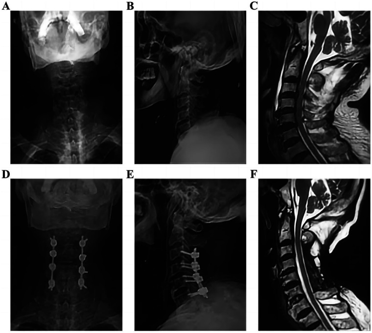
Multilevel stenosis with CCS before and after posterior decompression and fusion. 38 / 51~1 min · at 40:15Surgery & timingA brief slide for the surgical trainees; emergency colleagues can treat it as background.
The principle is to match the operation to the compression. Focal anterior compression from one or two disc-osteophyte levels is addressed anteriorly with discectomy or corpectomy and fusion; ACDF was the most common procedure in Brodell's national data Brodell 2015. Multilevel stenosis, especially with OPLL and a lordotic or neutral alignment, is addressed posteriorly by laminoplasty or laminectomy and fusion, as in the aside. Kyphosis reduces the drift-back that posterior decompression relies on, and may push you to an anterior or combined approach.
Instability, whether from fracture, dislocation or ligamentous disruption, mandates fusion, and dislocations are reduced promptly.
Aarabi's group confirmed decompression on postoperative MRI in every patient, and residual compression matters for recovery Aarabi 2021. Whatever you do, make sure the cord is actually free.
The comparative literature on approach is retrospective and reviewed in Divi's narrative review Divi 2019; there is no high-quality evidence that one approach is superior for the syndrome itself.
Refs: Brodell et al., Spine J, 2015 · Aarabi et al., J Neurotrauma, 2021 · Divi et al., Global Spine J, 2019- 39 / 51~1 min · at 41:15Surgery & timing
Twenty seconds for the audience.
Option C is correct. This is exactly the patient for whom the evidence is strongest: AIS C, persistent compression, stable physiology. Lenehan's consensus, the JAMA Surgery subgroup and the 2024 guideline all point the same way Lenehan 2010 Badhiwala 2022 Fehlings 2024.
Option A confuses stability with the surgical indication. Option B reflects 1970s teaching that no longer holds for this severity. Option D inverts the priorities: MAP support is delivered around surgery, not as a prerequisite to it.
Refs: Badhiwala et al., JAMA Surg, 2022 · Fehlings et al., Global Spine J, 2024 · Lenehan et al., Spine (Phila Pa 1976), 2010 - 40 / 51~0 min · at 41:25Prognosis
Section seven: prognosis, about five minutes.
- 41 / 51~1 min · at 42:40Prognosis
Recovery numbers for counselling.
In Dvorak's long-term follow-up from Vancouver, the mean ASIA motor score improved from 58.7 at injury to 92.3, 86% walked independently and 81% were continent, yet many remained significantly disabled and less functional than the general population, largely because of the hands Dvorak 2005. In Aarabi's stenotic surgical cohort the motor score went from 64 to 94 at one year, but manual dexterity was still only 64% of baseline and dysesthetic pain persisted Aarabi 2011.
Age drives function. In Penrod's series, 97% of patients under 50 walked independently at discharge versus 41% of those 50 and over Penrod 1990. Newey's natural history study found that all patients under 50 walked with bladder control at eight years while those over 70 did poorly and most had died Newey 2000.
Mortality is the sobering part. In the Welsh national cohort, nearly a third of central cord patients had died within five years, 56% of those over 70, and respiratory failure was the leading cause; the type of management did not affect mortality, age did Stanley 2023. This is a serious injury in an older person, whatever the motor score does.
- 42 / 51~1 min · at 43:55Prognosis
Predictors, for the conversation with the family.
On the favourable side: the admission motor score is the strongest predictor in every series, and AIS D patients do far better than AIS C Aarabi 2011 Pouw 2011 Dvorak 2005. Younger age, both for motor recovery and for function Penrod 1990 Ronzi 2018. The MRI: Ishida's series of upper-extremity-only injuries treated nonoperatively found that absence of cord signal change was the strongest predictor of full recovery, with an odds ratio of 64 Ishida 2002, and Wilson's prediction model added absence of edema or hemorrhage to AIS grade, motor score and age Wilson 2012. Merriam's clinical predictors, good hand function, hyperpathia and normal perianal sensation, still hold Merriam 1986. And the systemic factors: fewer comorbidities, and the absence of spasticity at follow-up Dvorak 2005.
The recovery pattern is the one Schneider described: the legs recover first, then bladder function, then the proximal arms, with the hands last and least Schneider 1958. Most of the gain happens in the first weeks to months. The long-term complaints are dysesthetic hand pain and spasticity, not walking.
And a reminder from the prospective European cohort: the AIS grade predicts function better than the central cord label does Pouw 2011. When you counsel, counsel from the grade.
Refs: Aarabi et al., J Neurosurg Spine, 2011 · Pouw et al., Spinal Cord, 2011 · Dvorak et al., Spine (Phila Pa 1976), 2005 · Penrod et al., Arch Phys Med Rehabil, 1990 · Ronzi et al., Ann Phys Rehabil Med, 2018 · Ishida & Tominaga, Spine (Phila Pa 1976), 2002 · Wilson et al., J Neurotrauma, 2012 · Merriam et al., J Bone Joint Surg Br, 1986 · Schneider et al., J Neurol Neurosurg Psychiatry, 1958 - 43 / 51~1 min · at 44:40Prognosis
Complications are common and mostly medical.
In the NACTN prospective cohort of 315 acute SCI patients, 58% had at least one complication, most within two weeks; the rate rose with severity, from 25% in AIS D to 84% in AIS A. Respiratory failure and pneumonia led the list, and mortality was 3.5% Grossman 2012.
Respiratory complications are the killers in the older central cord population, as the Welsh data showed Stanley 2023. Aspiration precautions, chest physiotherapy, early mobilisation and a low threshold for a controlled airway are the defence.
Venous thromboembolism: prophylaxis within 72 hours per the guideline Dhall 2013; the 2024 meta-analysis found fewer thromboembolic events with early surgery, presumably through earlier mobilisation Sattari 2024.
Bladder and bowel, pressure areas, delirium and deconditioning are the routine hazards of an immobilised older patient and need the same vigilance as after a hip fracture.
Neuropathic pain, spasticity and the psychological impact of losing hand function are the long-term burdens; involve rehabilitation medicine early.
And, as discussed, the treatment itself has complications: cardiac events with vasopressors in older patients Readdy 2015.
Refs: Grossman et al., J Neurosurg Spine, 2012 · Stanley et al., Bone Joint J, 2023 · Dhall et al., Neurosurgery, 2013 · Sattari et al., Spine J, 2024 · Readdy et al., J Neurosurg Spine, 2015 - 44 / 51~0 min · at 44:50Cases
Section eight: two cases, about eight minutes. Make the audience commit to an answer at each step before revealing the next.
The 72-year-old from the first slide
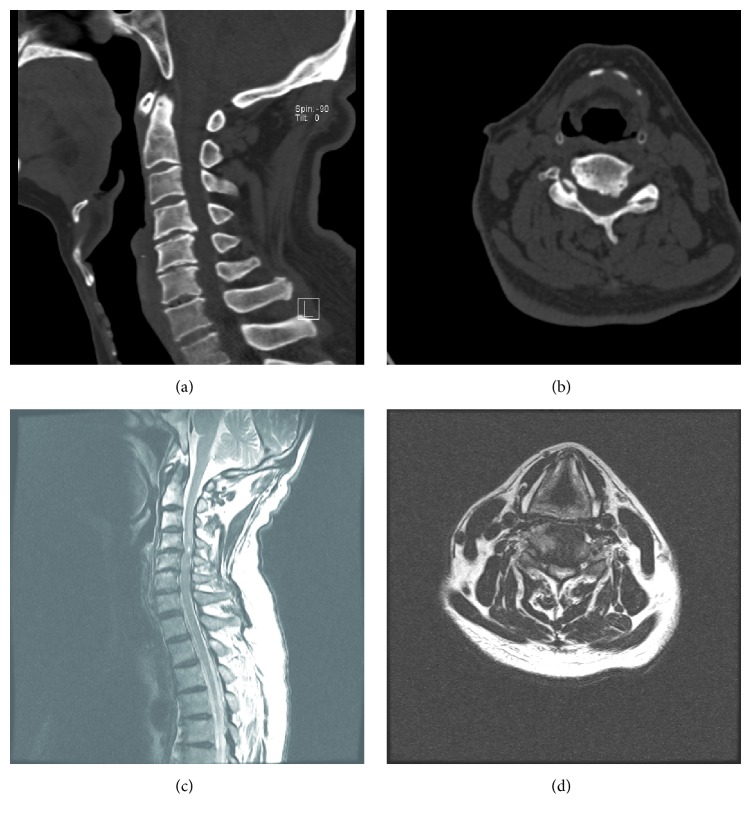
Delayed CCS after fall, 69-year-old man: CT and MRI 45 / 51~4 min · at 48:20CasesCase one is the patient from the first slide. Walk through it step by step and make the audience commit at each question.
Stage one: the examination. He is bradycardic and his blood pressure is low-normal, which in a hypertensive man is a relative hypotension. The motor scores show a 22-point gap, the reflexes in the legs are brisk, the sacral examination is preserved and the bladder is retaining. Ask: what is the diagnosis and the grade?
Stage two: an incomplete cervical cord injury with a central cord pattern, AIS D because most of his key muscles are grade 3 or better. Point out the mechanism clue, the forehead, and that the bradycardia may be neurogenic, beta-blockade, or both. Ask about imaging.
Stage three: the CT. Spondylosis, disc-osteophyte complexes at C4-5 and C5-6, no fracture. This is exactly the report that leads to a missed diagnosis. He still has a deficit, so the CT does not end the workup. Ask what is next.
Stage four: the MRI. Compression at C4-5 with cord signal, no hemorrhage, no ligamentous injury. The spine is stable; the cord is compressed. Note the image shows a comparable case of C4-5 osteophyte compression with intramedullary signal, from an open-access report.
Stage five: the next hour. Collar and no extension; catheter for the retention; arterial line and a MAP floor of 75 to 80 with fluid and then norepinephrine, avoiding dopamine at his age Kwon 2024 Readdy 2015. Hold the apixaban and discuss reversal, because surgery is on the table. No steroids: he is AIS D, older, diabetic, and the evidence does not support it Fehlings 2017. Call the surgeon now, at two and a half hours from injury, not in the morning. ICU, hourly checks, mechanical VTE prophylaxis.
Stage six: the decision. This is the genuinely debatable case. AIS D with an improving examination can be observed Lenehan 2010, and the propensity-matched cohort found no benefit of early surgery in AIS D Badhiwala 2022. But he has severe persistent compression at a level with cord signal, he is diabetic and at risk of a second fall, and the 2024 guideline frames early surgery as the preferred option across SCI Fehlings 2024. A reasonable surgeon might observe for 24 to 48 hours; another might decompress within 24 hours once anticoagulation is reversed. In this version he had an ACDF at 19 hours and did well, with the typical residual hand dysesthesia. Emphasise that the emergency department's job was done correctly whichever way the surgeon chose: the diagnosis was made, the cord was protected and perfused, the consult was early, and the anticoagulation problem was already being solved.
Pearl: The emergency department cannot make the timing decision, but it can make it possible: early recognition, early MRI, early consult, early reversal of anticoagulation.A 24-year-old after a dive
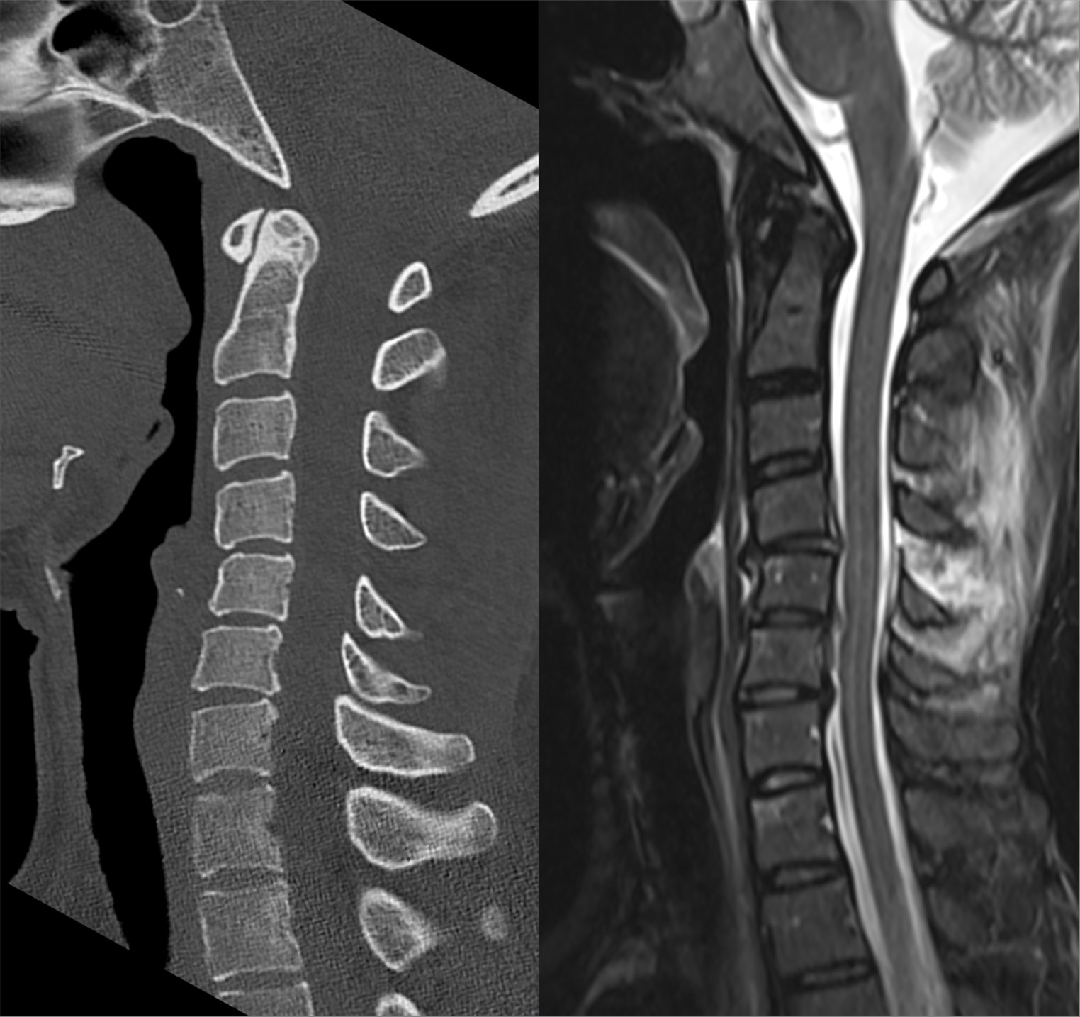
Hyperflexion injury C4/C5: CT and T2 fat-sat MRI 46 / 51~3 min · at 50:50CasesCase two is the younger, high-energy version.
Stage one: hypotension with bradycardia and warm peripheries in a young man with a negative FAST scan. The motor scores show profound upper limb weakness with legs at grade 3, and sacral sparing.
Stage two: this is neurogenic shock, and the treatment is volume then a vasopressor with chronotropic effect, with atropine available for bradycardia Ruiz 2018. The grade is AIS C. With this mechanism, expect a structural injury.
Stage three: the CT shows a C6-7 fracture-dislocation. The spine is unstable. The image is a comparable open-licensed CT of a C6-7 fracture-dislocation. Ask the room whether he needs an MRI before reduction.
Stage four: the honest answer is that practice varies. In an awake, examinable patient many surgeons proceed to closed reduction with serial examinations. An MRI before reduction can identify a disc herniation that might be driven into the cord during reduction, and in most modern centres it can be obtained without meaningful delay, so it is usually done. Here it shows an extruded disc behind C6, cord edema across three levels and a disrupted posterior ligamentous complex: a surgical injury from every angle. The image on the slide is a comparable open-licensed case: subtle interspinous widening on CT and, on the fat-saturated MRI, extensive edema through the posterior ligamentous complex, the injury the CT understates. The plan is anterior discectomy to clear the disc, open reduction and fusion, with posterior stabilisation as needed.
Stage five: how urgent? This is the patient for whom the timing evidence is least ambiguous: young, AIS C, structural compression from a disc and dislocation, the group that gained most in Guest's series and in the propensity-matched cohort Guest 2002 Badhiwala 2022. Surgery at 7 hours, after resuscitation, is the target. MAP support in the guideline range for several days Kwon 2024; no steroids at this centre. He developed pneumonia, the commonest complication Grossman 2012, and recovered to AIS D with persisting hand weakness.
Contrast the two cases explicitly: same syndrome, opposite structural problems, and a timing decision that was debatable in the first and clear in the second.
- 47 / 51~1 min · at 51:35Cases
Quick question to close the cases. Option C. She is outside the eight-hour window, both guidelines advise against steroids at that point, the 48-hour regimen is never recommended, and her age raises the harms Hurlbert 2013 Fehlings 2017 Evaniew 2016. MRI findings do not change the steroid decision.
Refs: Hurlbert et al., Neurosurgery, 2013 · Fehlings et al., Global Spine J, 2017 · Evaniew et al., J Neurotrauma, 2016 - 48 / 51~1 min · at 52:50Summary
Six take-home points. Read them slowly; this is the slide people photograph.
One: the pattern. Two: the mechanism and why the CT is normal. Three: the imaging rule and the documentation that makes a consult useful. Four: the medical management, with the updated MAP target and the steroid position. Five: the consult and the timing evidence, with its strength stated honestly. Six: the counselling.
If the audience remembers only the first and third points, patients will still be better off.
- 49 / 51~1 min · at 53:20Summary
Close on the tension that runs through the whole talk. The biology and the modern evidence say that decompression is time-sensitive. The epidemiology says that the typical patient is old, frail, anticoagulated and at risk from the treatment itself. Good care lives in the space between those two truths, and it starts in the emergency department.
Thank the audience and open for questions.
- 50 / 51~0 min · at 53:30Summary
Selected references. The complete list of 95 verified sources, with PubMed and DOI links, is on the references page of the lecture site; the QR code on the next slide takes the audience there.
- 51 / 51~0 min · at 53:45Summary
Leave this slide up during questions. The link opens the deck page with the complete, filterable reference list and the notes handout.